Microscopy Images by Camera
View sample Images by INFINITY Camera
INFINITYHD Sample Images
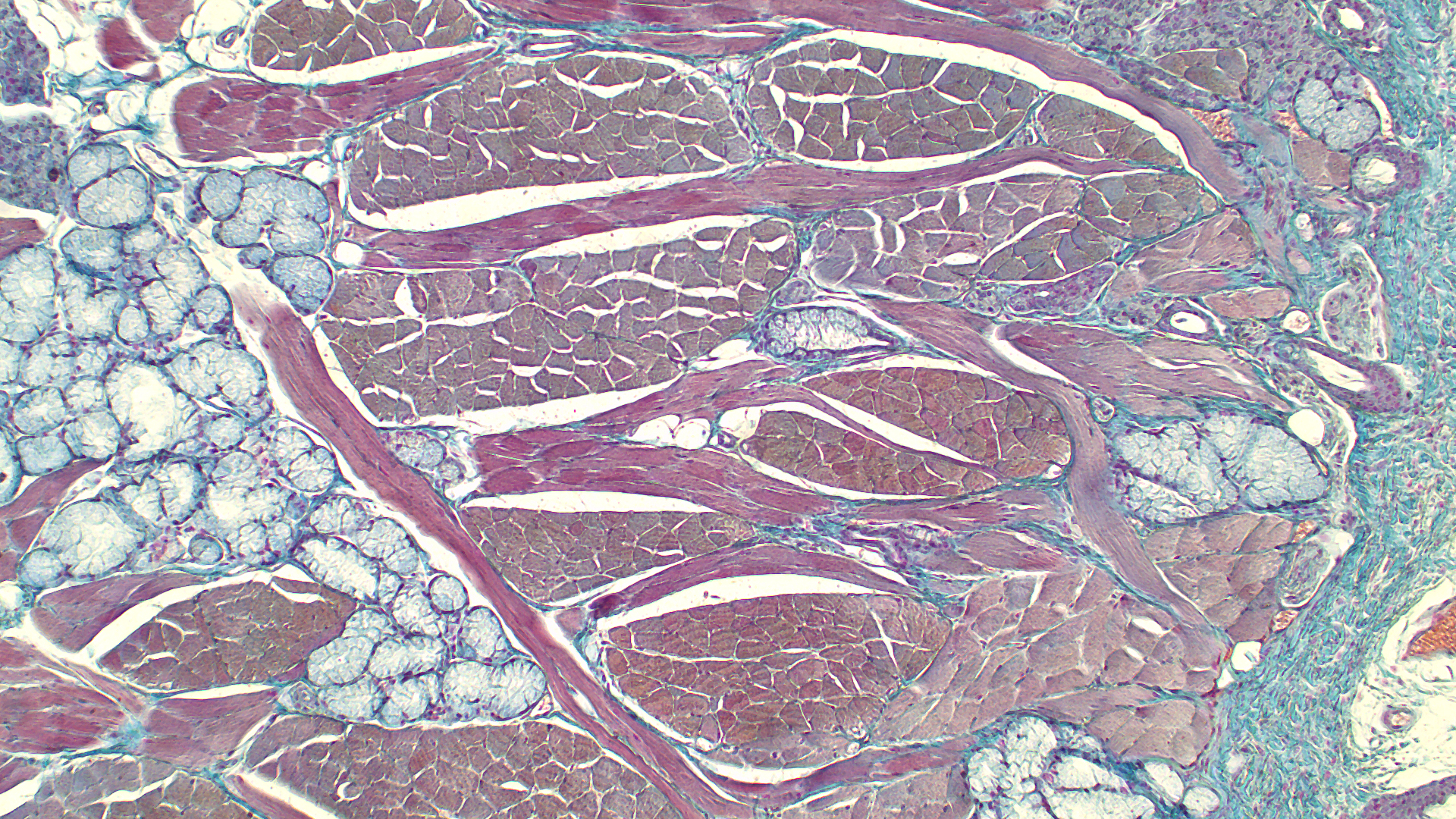
Rabbit Tongue
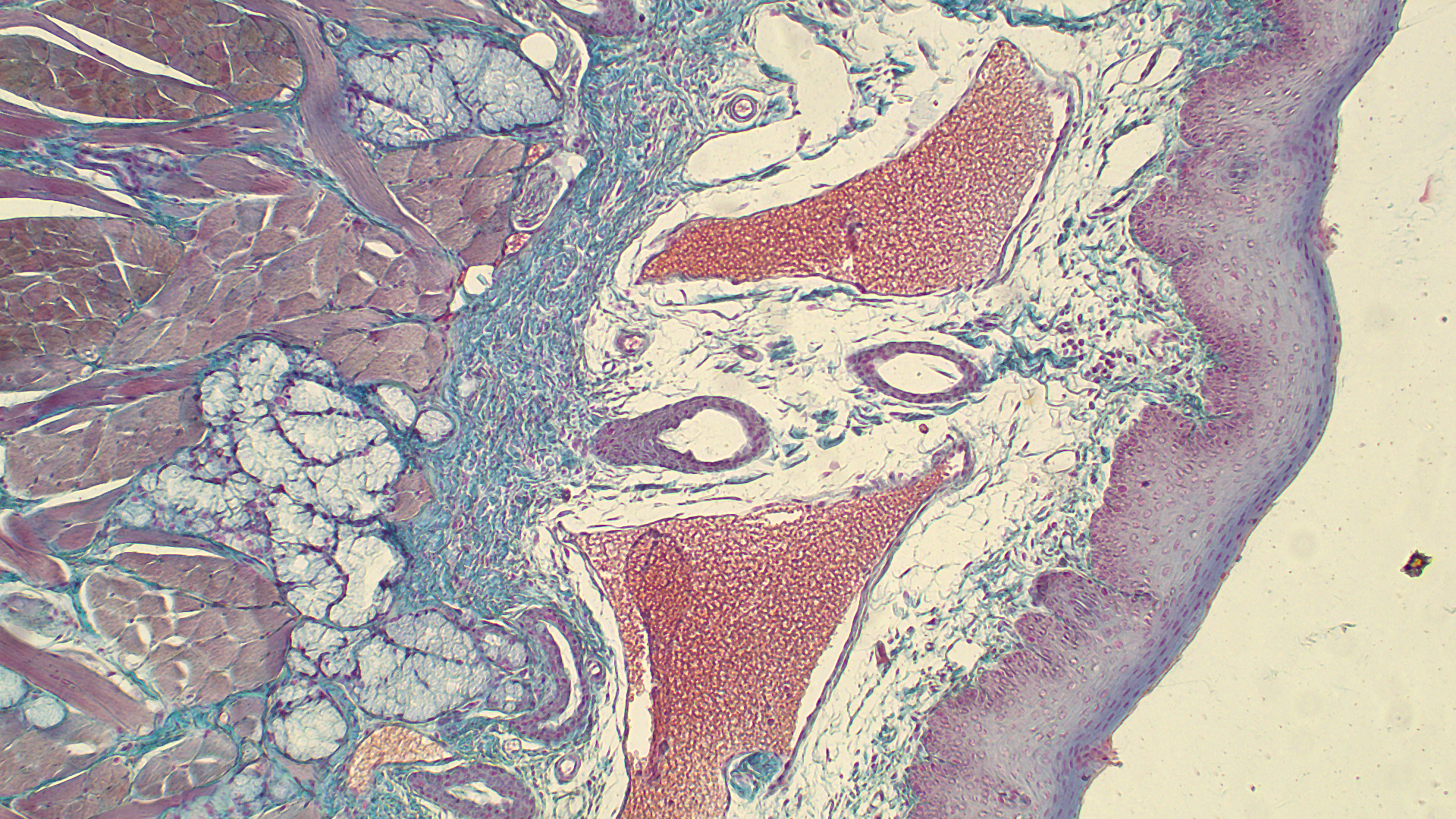
Rabbit Tongue
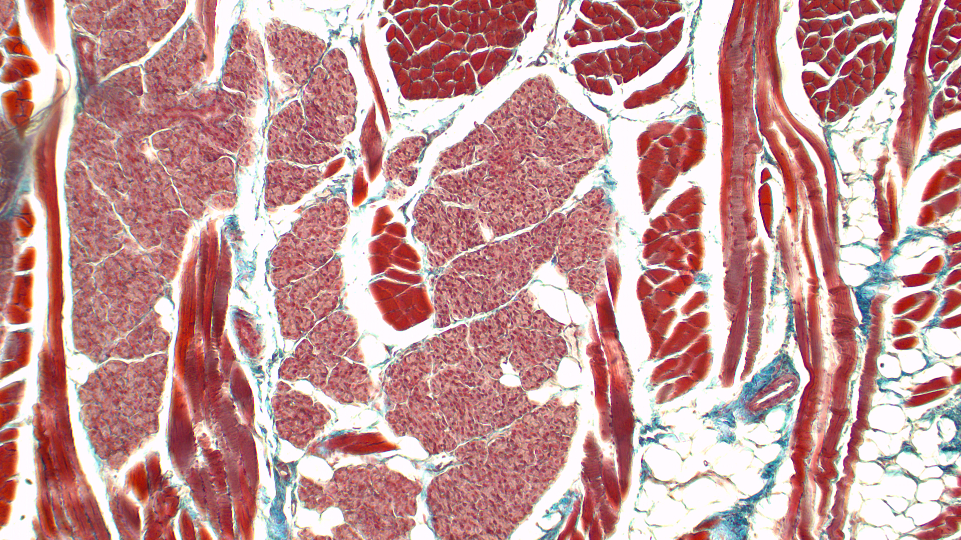
Rabbit Tongue
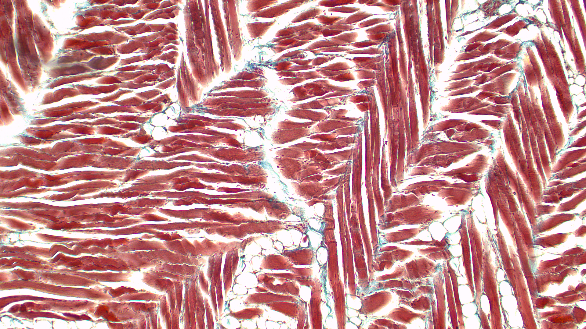
Rabbit Tongue
INFINITY1 Sample Images
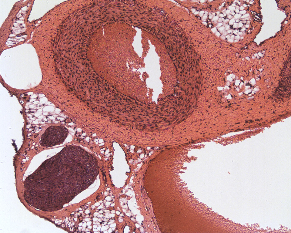
Arteries and Vein
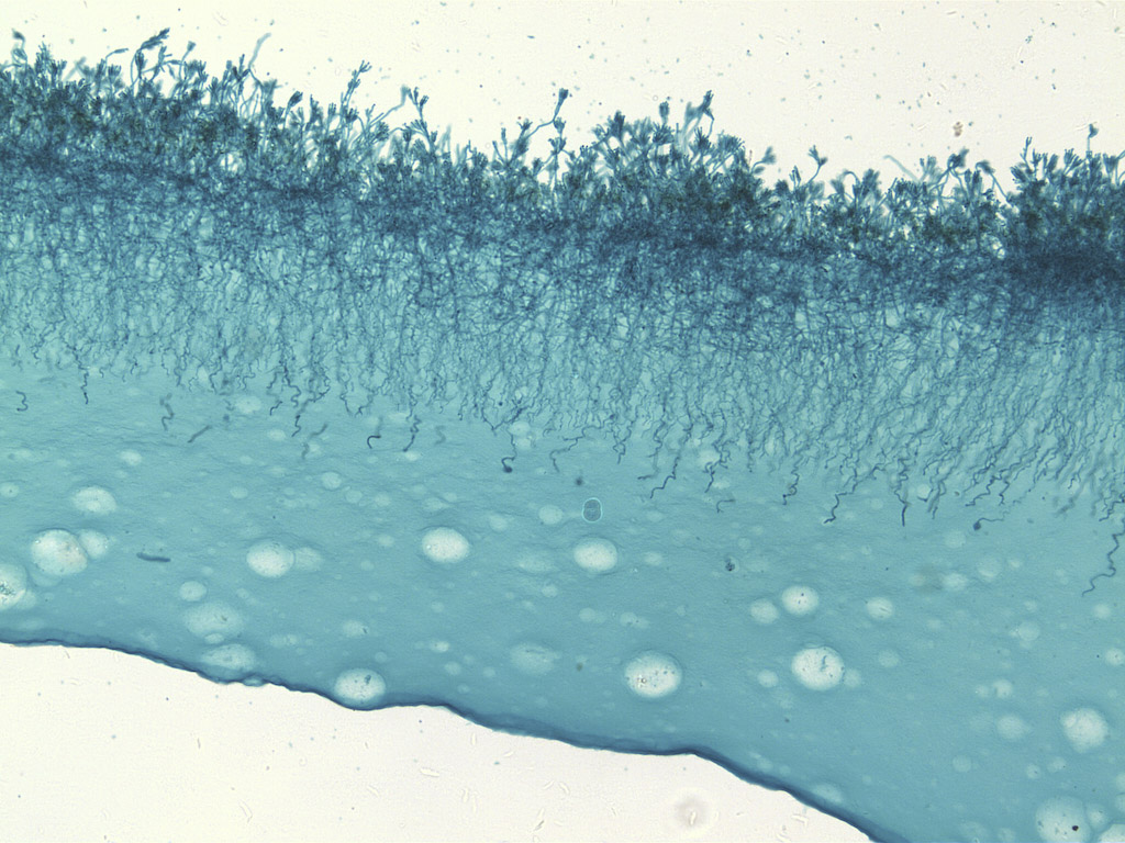
Penicillium

Stained Tissue
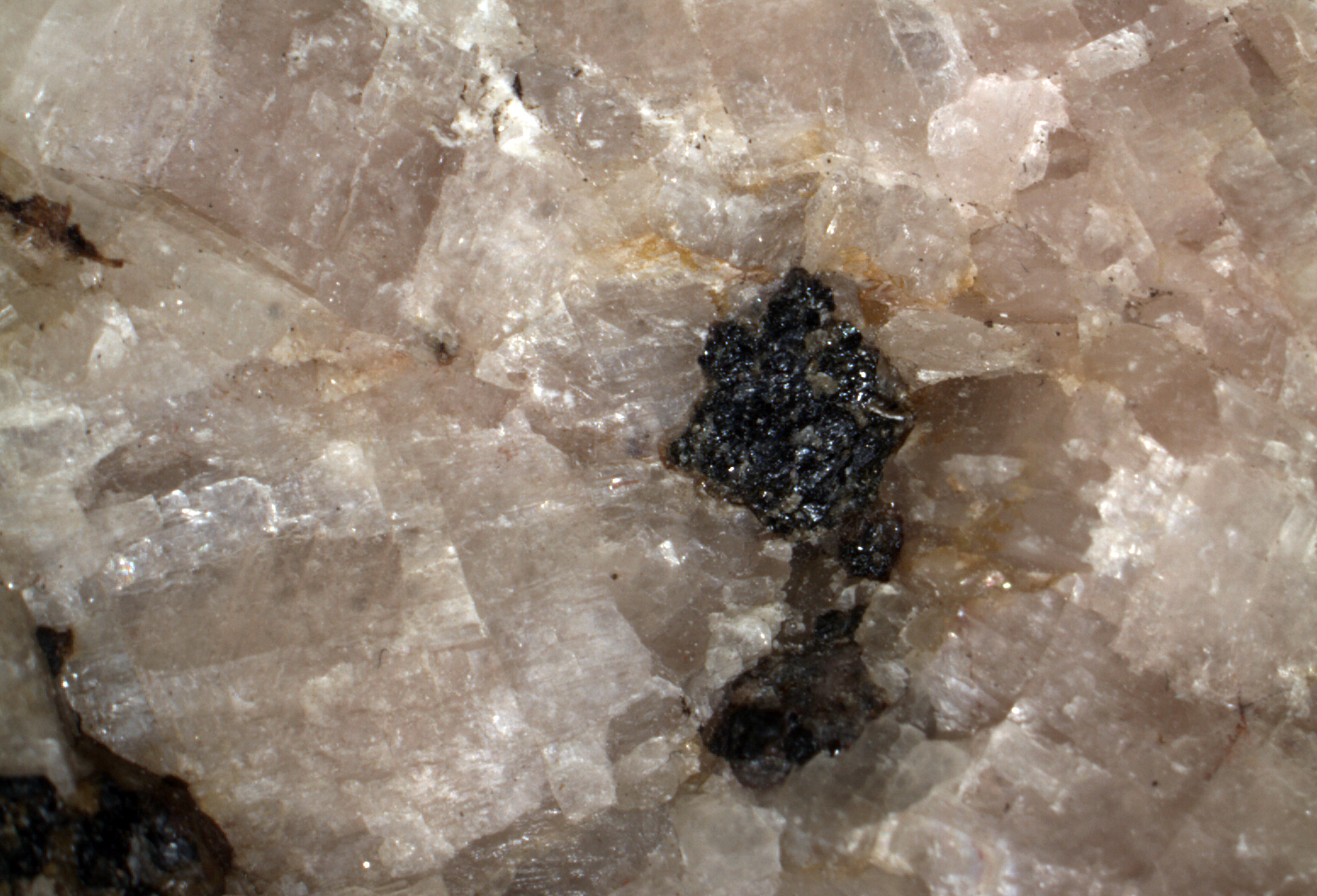
Mineral
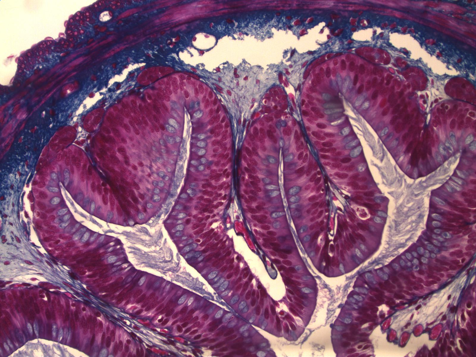
Columnar Epithelium
Corn Grain

Mammal Bone Marrow
Mammal Fundic Stomach
Concrete Specimen
INFINITY2-1 Sample Images
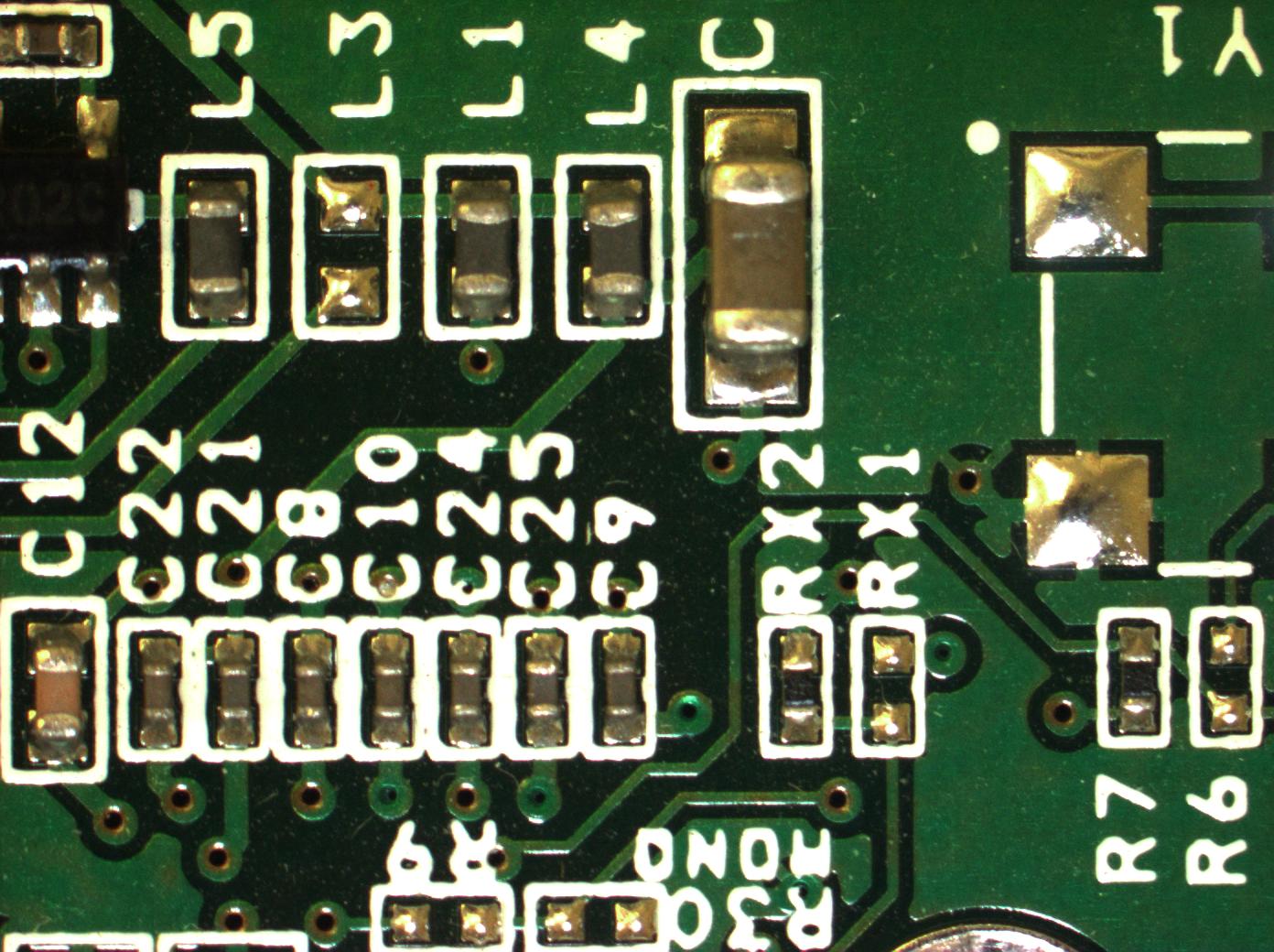
Circuit Board

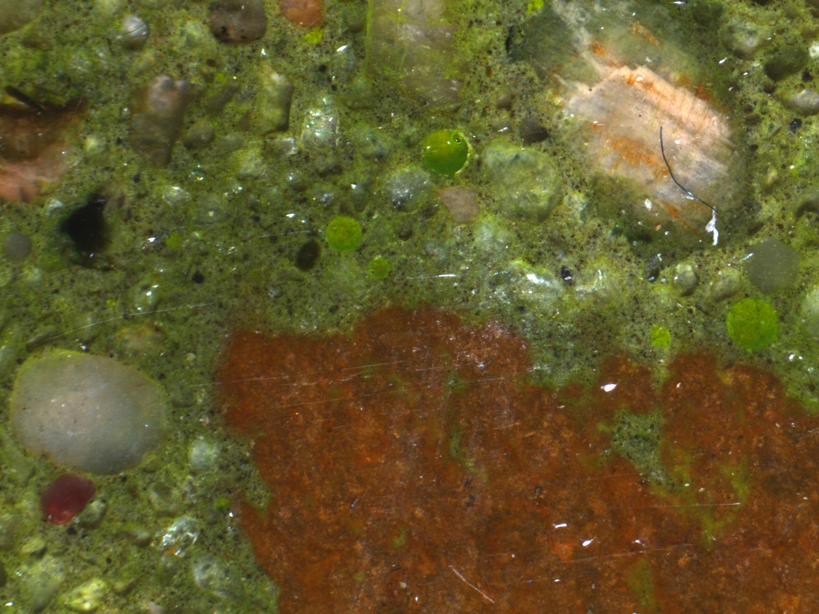
Rock Sample
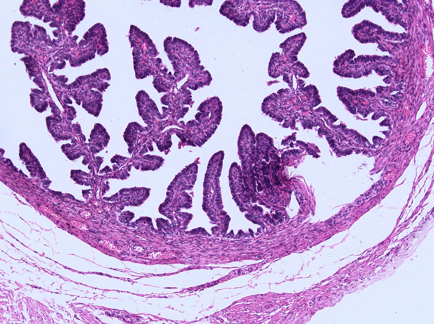
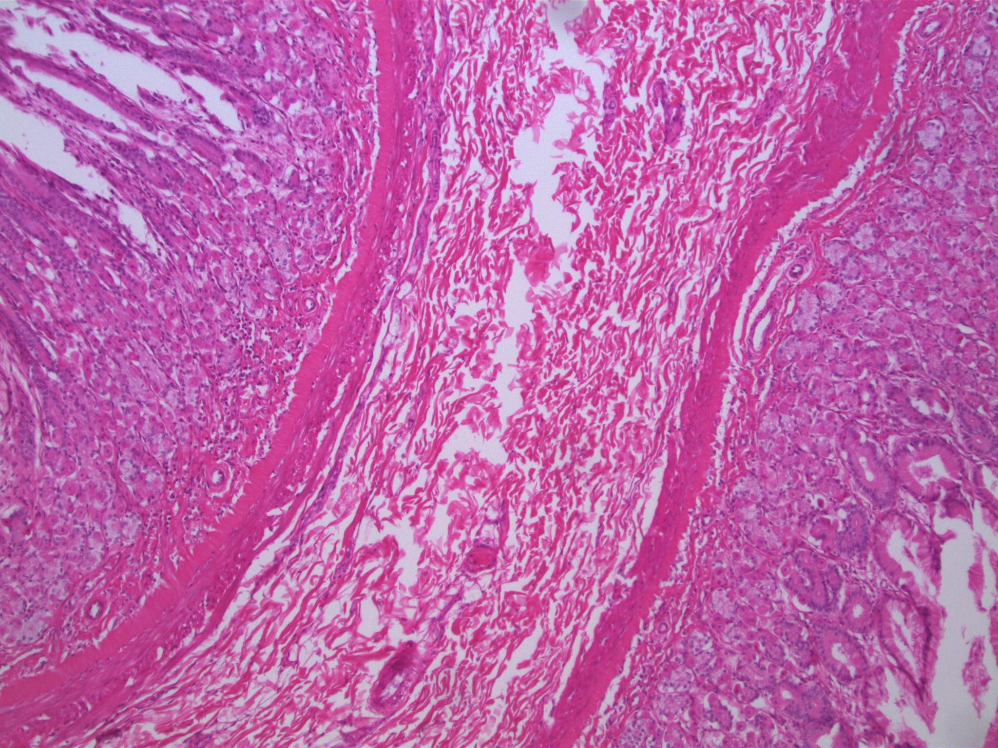
Colimnar Epithelium
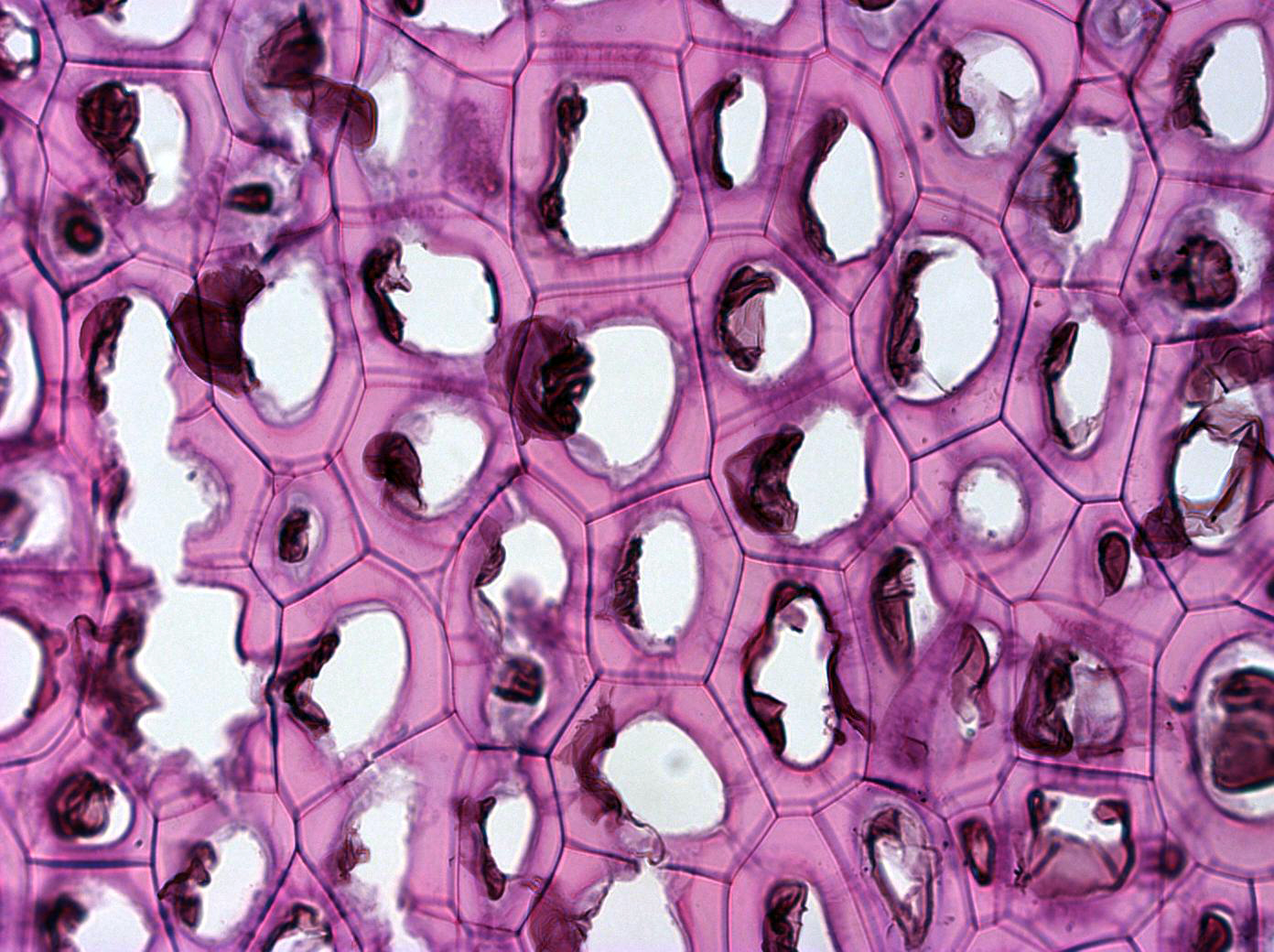
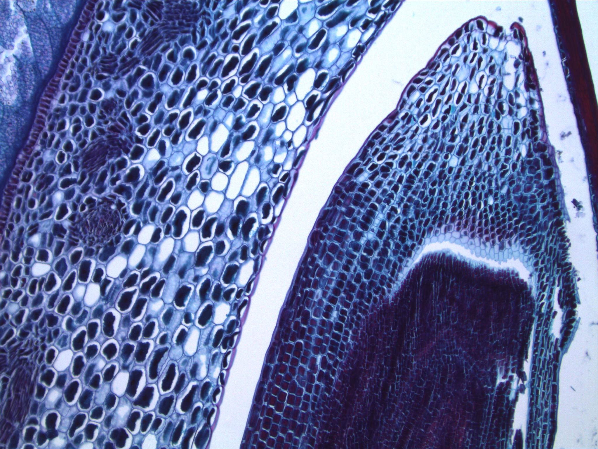
Persimmon Endosperm
.jpg)
Platinum nanoelectrode as it is being submerged into an aqueous solution
Photo taken by Netzahualcóyotl Arroyo Currás (PhD), Postdoctoral Scholar, Plaxco Group, University of California, Santa Barbara. Submitted in the Art of Science Competition.
INFINITY2-2 Sample Images
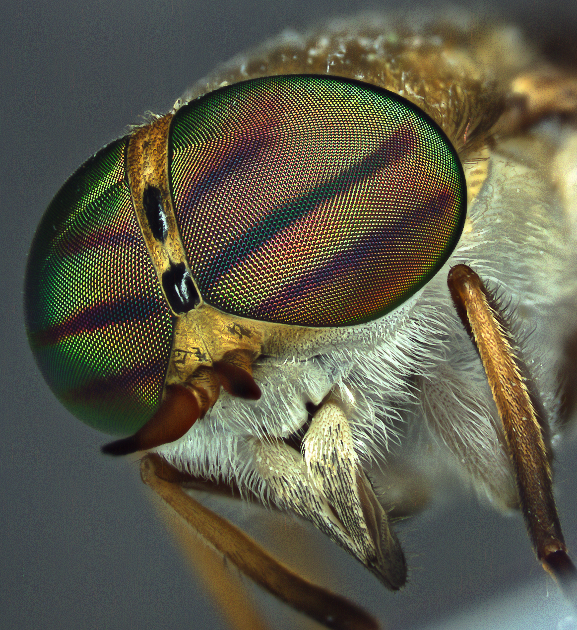
Biting Fly, Tabanidae, Diptera (Photo: William Fisher)
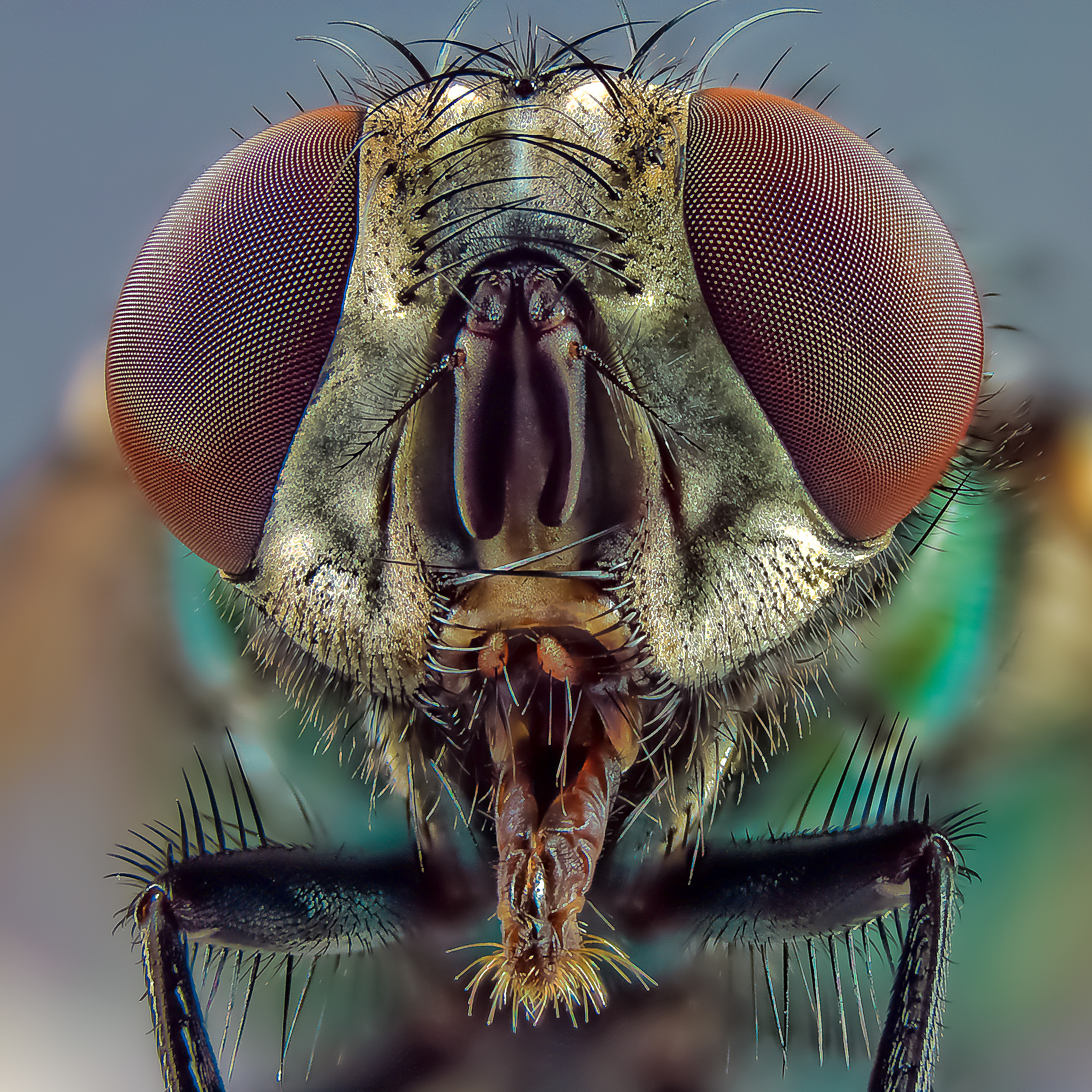
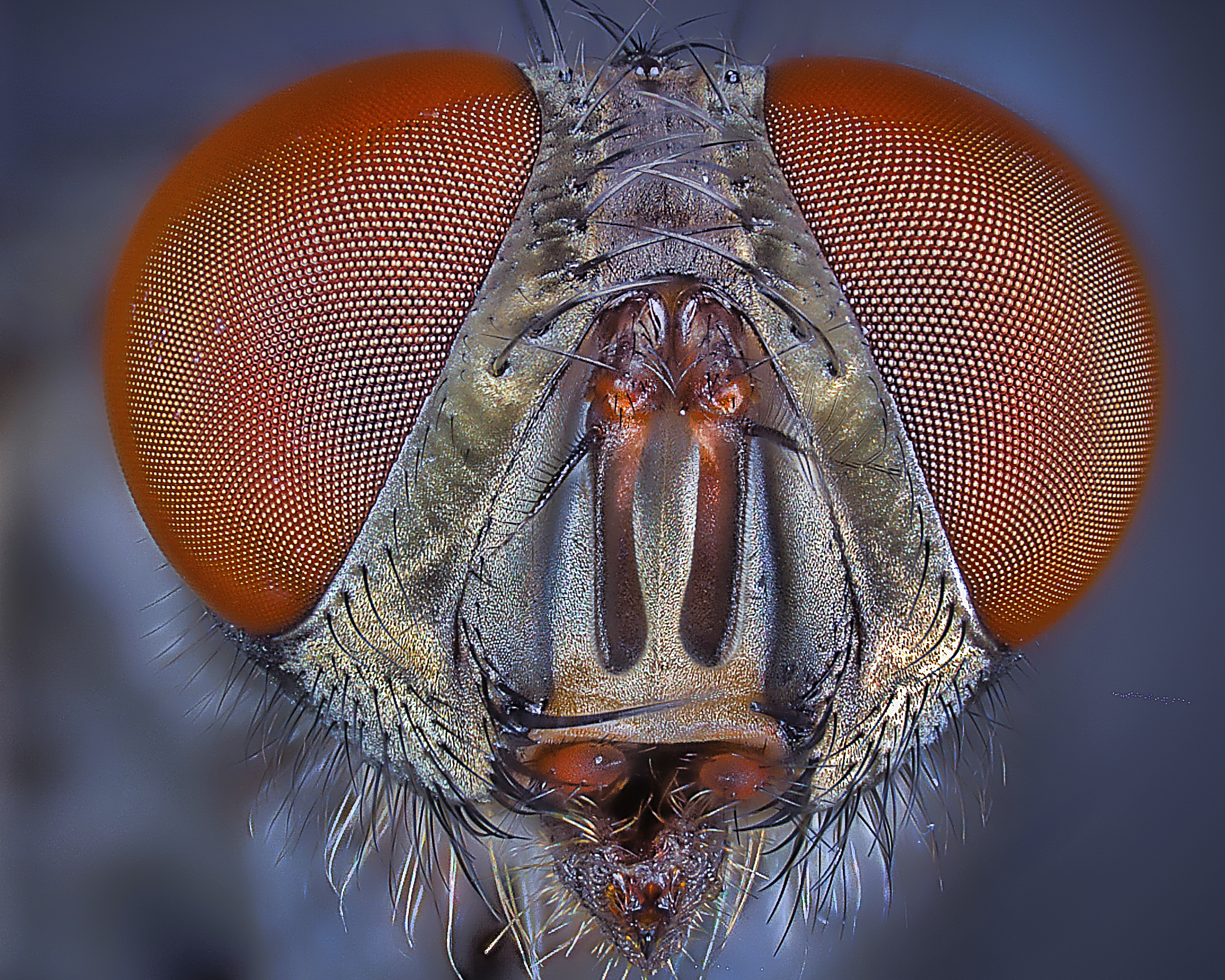
Blow Fly, Calliphoridae, Diptera (Photo: William Fisher)

Broad-Headed Sharpshooter, Oncometopia Orbana, Hemiptera (Photo: William Fisher)
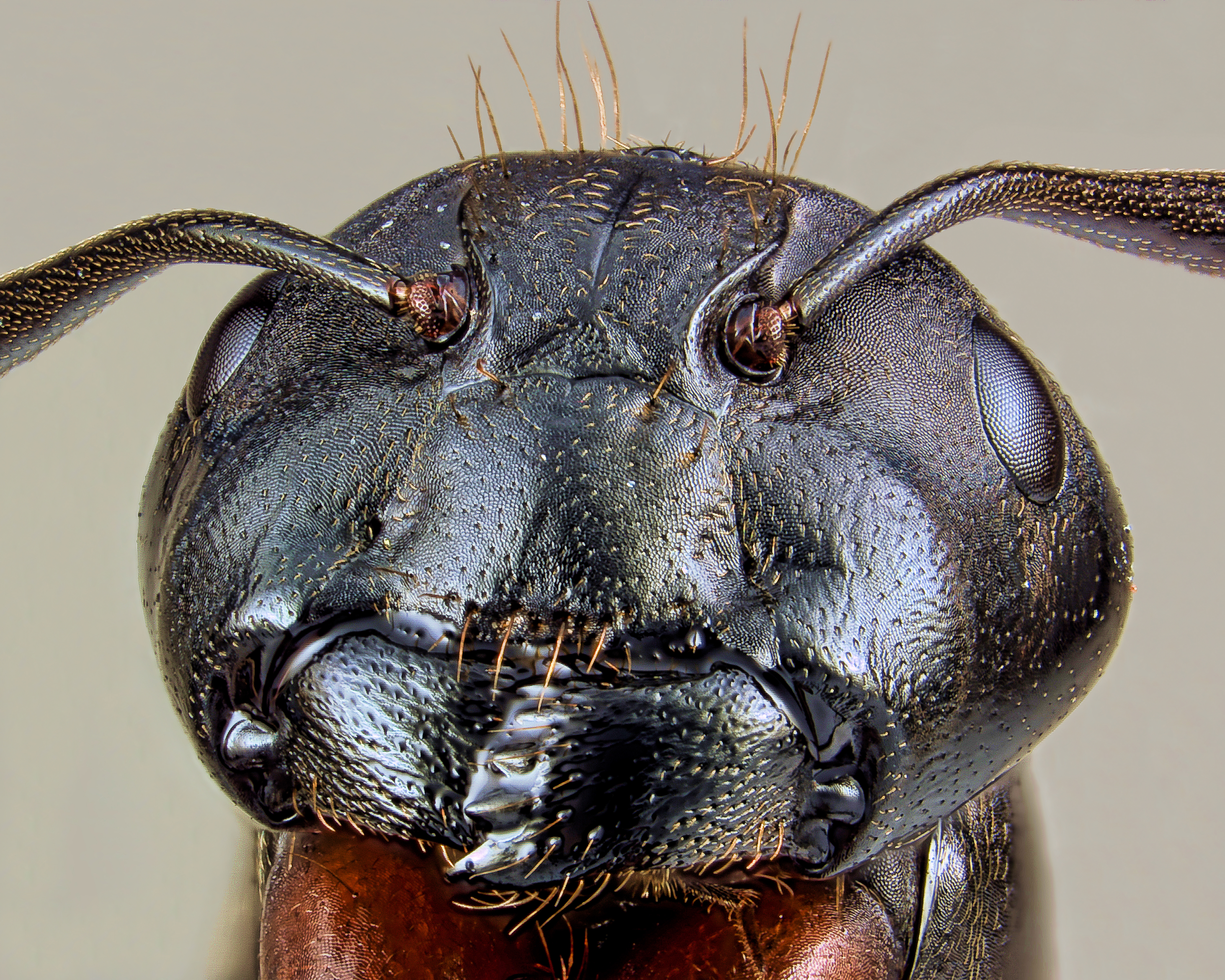
Carpenter Ant, Formicidae, Hymenoptera (Photo: William Fisher)
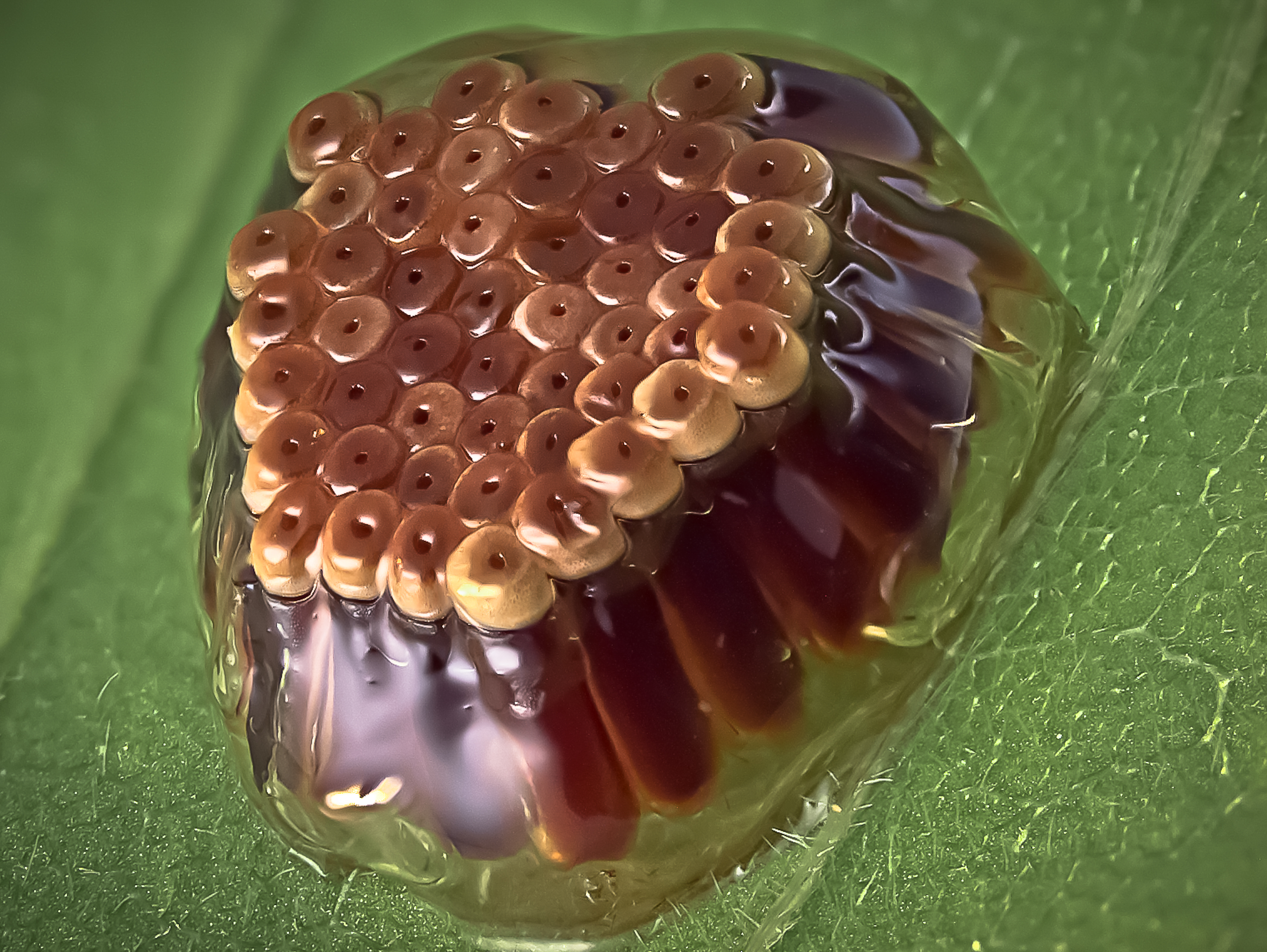
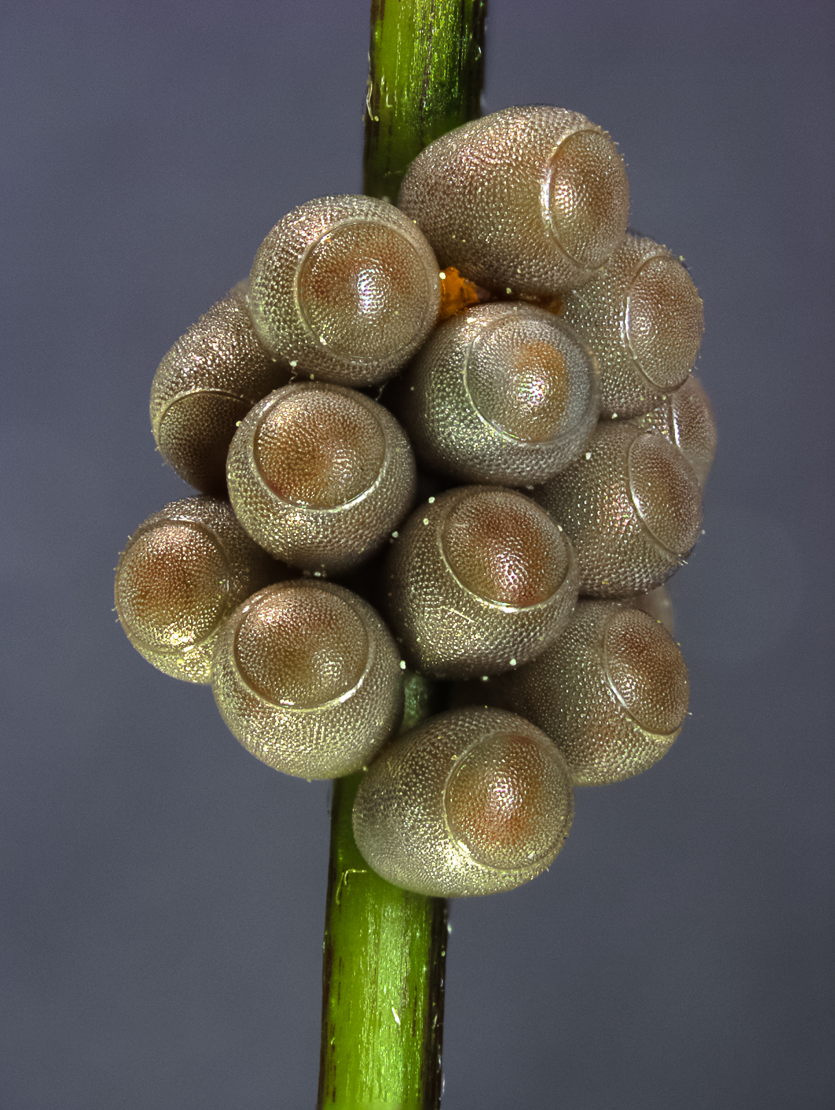
Egg Mass, Unknown Species, Hemiptera (Photo: William Fisher)
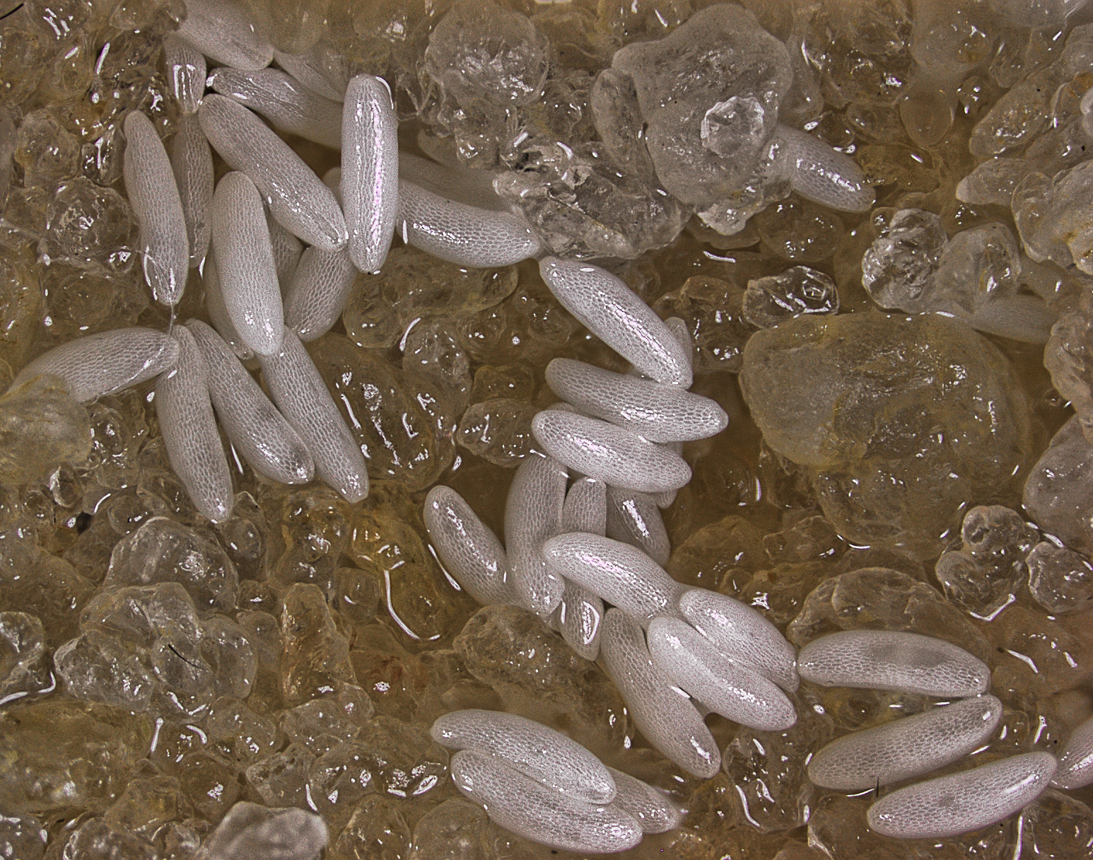
Eggs of Seedcorn Maggot, Delia Platura, Anthomyiidae, Diptera (Photo: William Fisher)
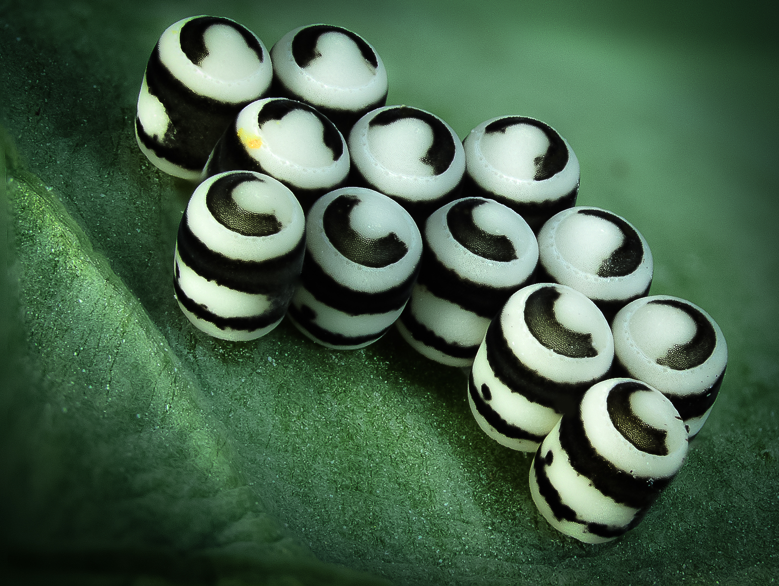
Eggs of the Harlequin Bug, Murgantia Histrionica, Hemiptera (Photo: William Fisher)

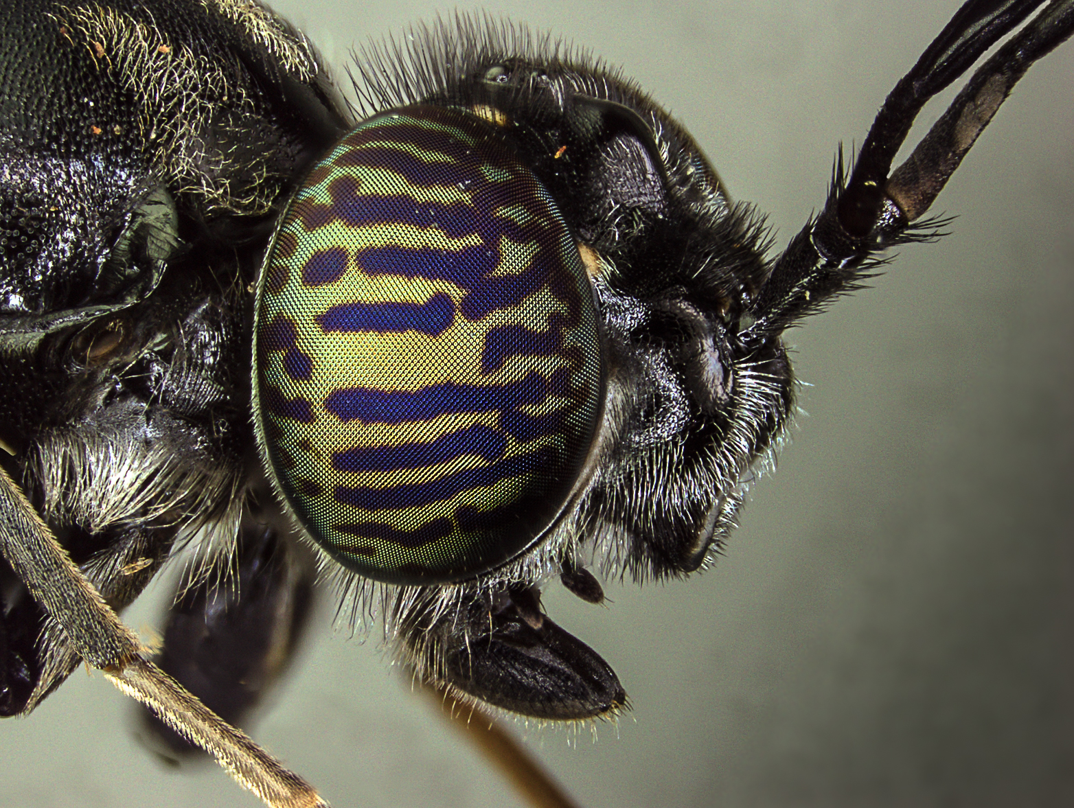
Eye of a Cicada, Hemiptera (Photo: William Fisher)

Greenhead Fly, Tabanus Nigrovittatus, Tabanidae, Diptera (Photo: William Fisher)
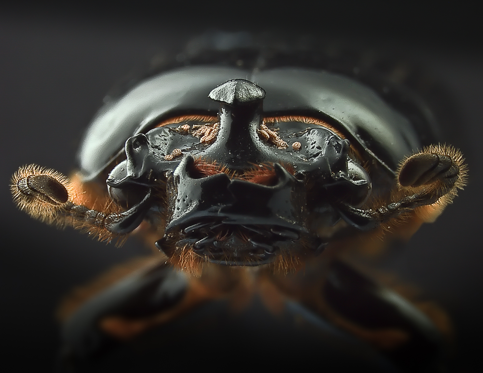
Horned passalus, Odontotaenius disjunctus, Passalidae, Coleoptera (Photo: William Fisher)
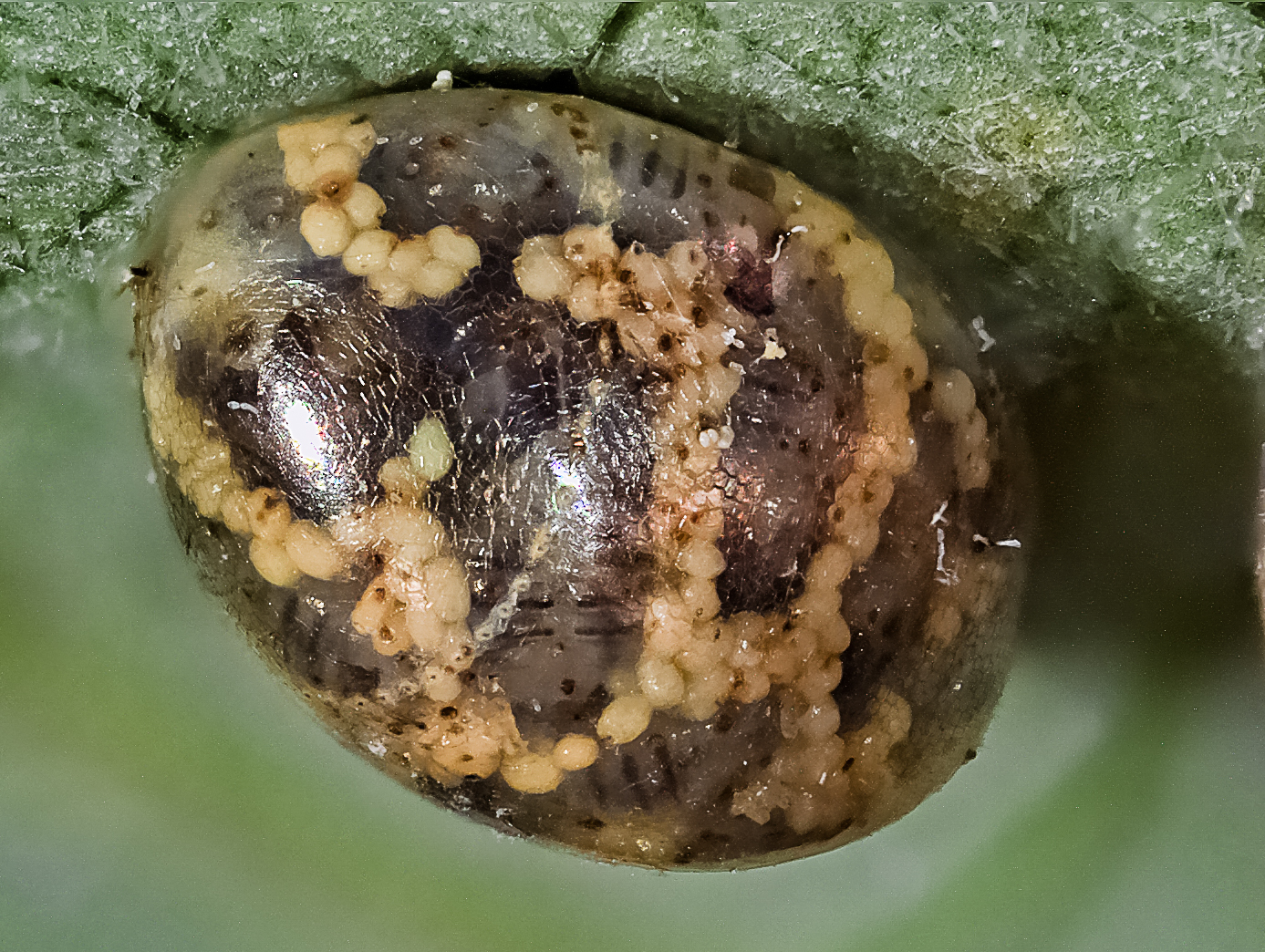
Insect Egg, Likely Parasitized (Photo: William Fisher)

Insect Eggs (Photo: William Fisher)
Tobacco Budworm egg, Heliothis Virescens, Noctuidae, Lepidoptera (Photo: William Fisher)
Unknown Fly 1, Diptera (Photo: William Fisher)

Unknown Fly 2, Diptera (Photo: William Fisher)

Unknown Fly 3, Diptera (Photo: William Fisher)
Unknown Fly 4, Diptera (Photo: William Fisher)
Unknown Insect Eggs (Photo: William Fisher)
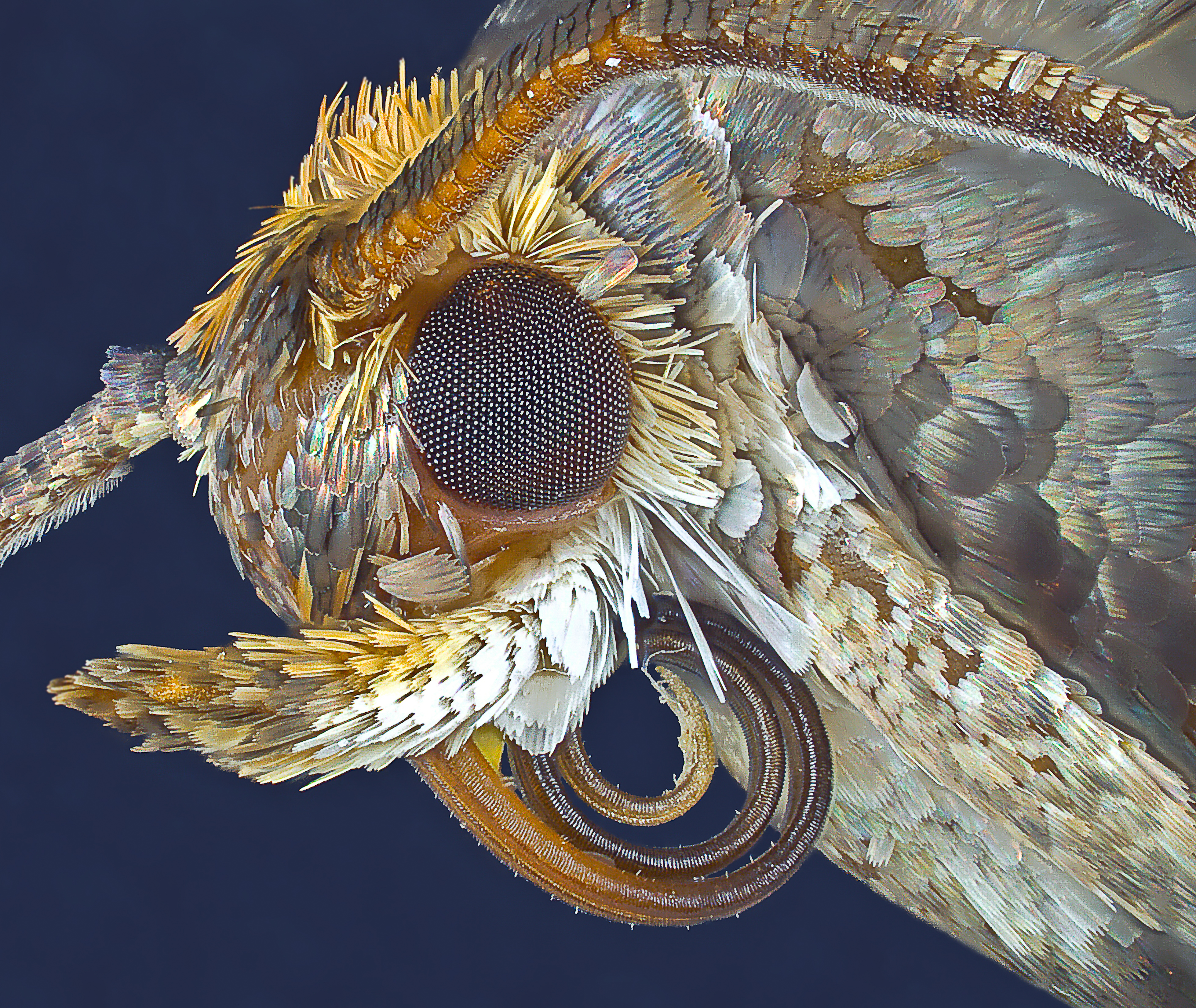
Unknown Moth, Lepidoptera (Photo: William Fisher)
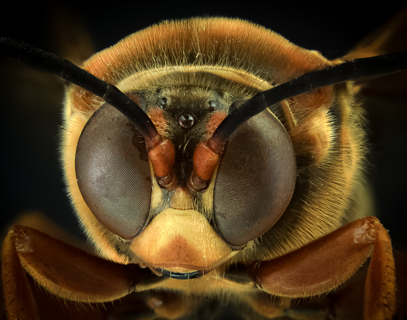
Unknown Wasp, Frontal, Hymenoptera (Photo: William Fisher)
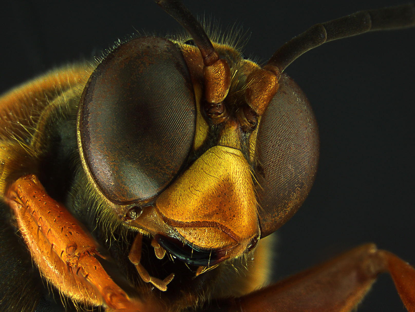
Unknown Wasp, Left Frontal, Hymenoptera (Photo: William Fisher)
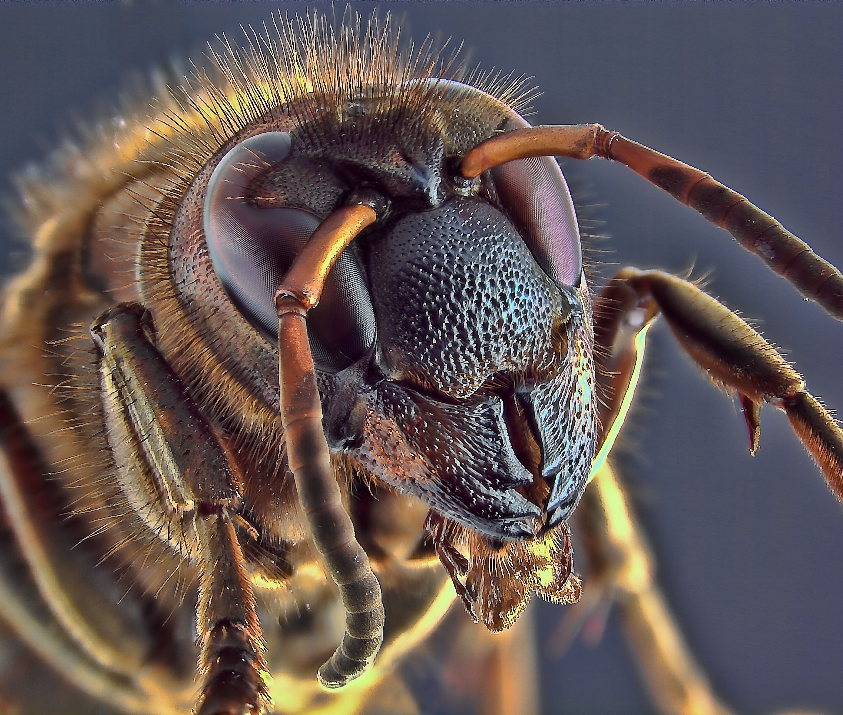
Wasp, Hymenoptera (Photo: William Fisher)
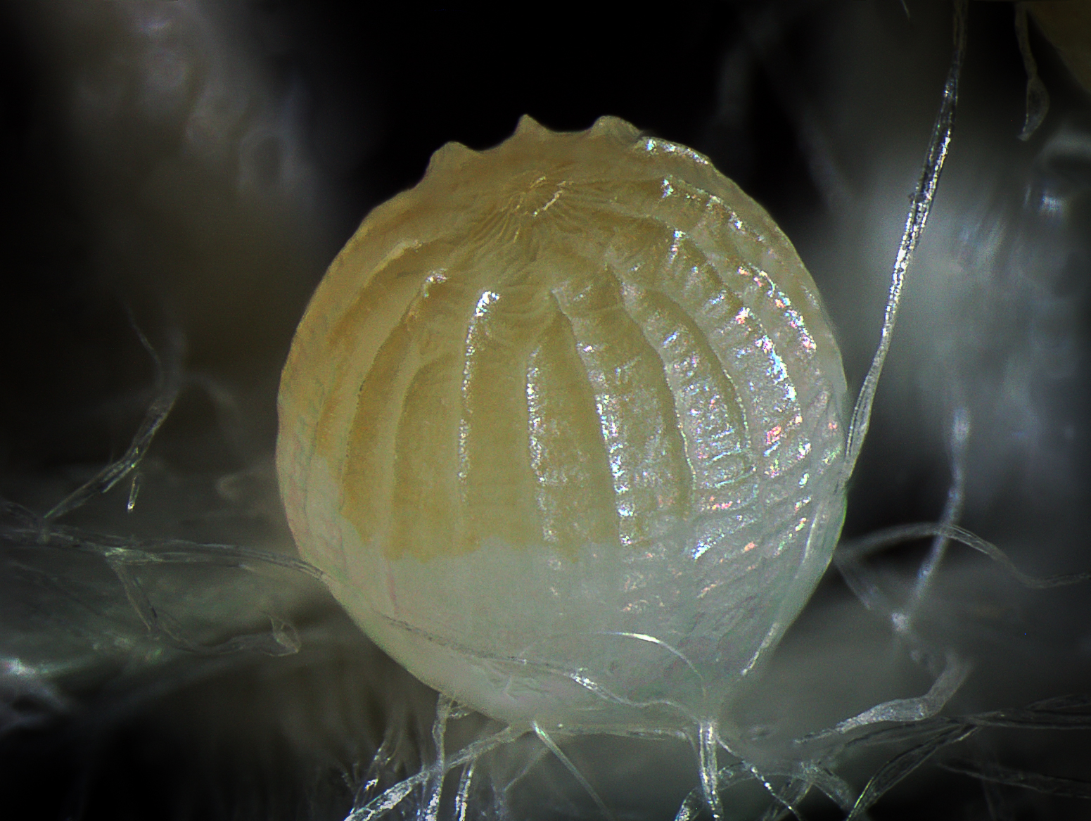
Western Flower Thrips Egg, Frankliniella Occidentalis, Thysanoptera (Photo: William Fisher)
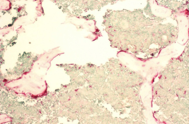
10x Bone Marrow - Femur TRAP Staining Osteoclast
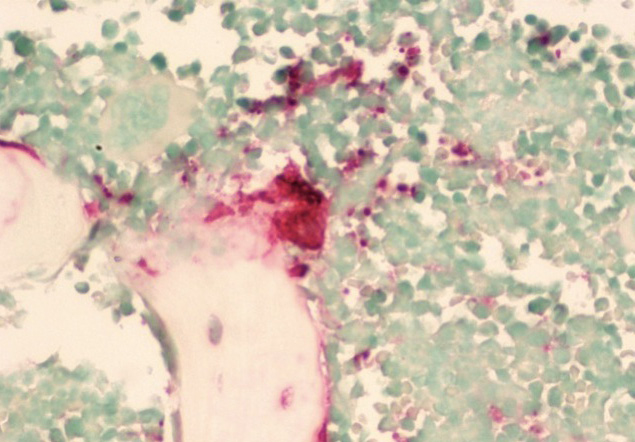
40x Bone Marrow - Femur TRAP Staining Osteoclast
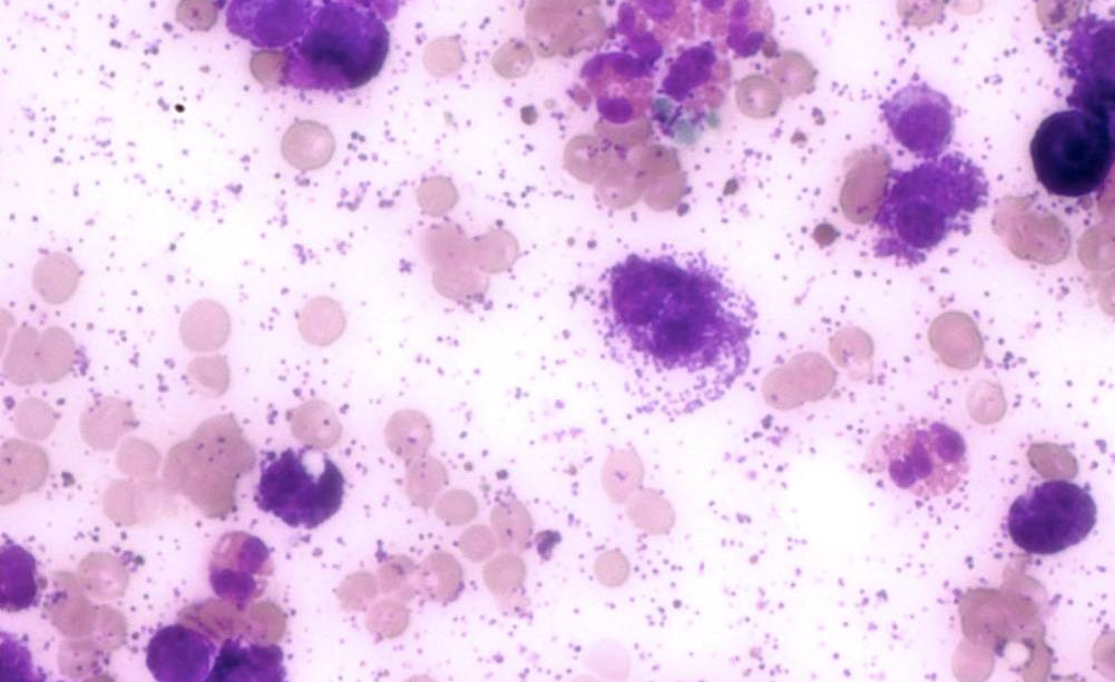
Canine Mast Cell Tumor Toluidine Blue Staining
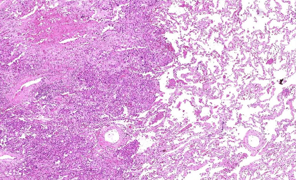
Lung Carcinoma - Human

Prostate Carcinoma - Human

Skin - Mouse
INFINITY2-5 Sample Images

Cellular Neurothekeoma 100x
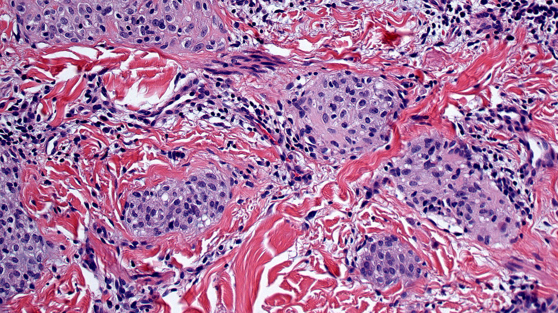
Cellular Neurothekeoma 200x
EPS 200x
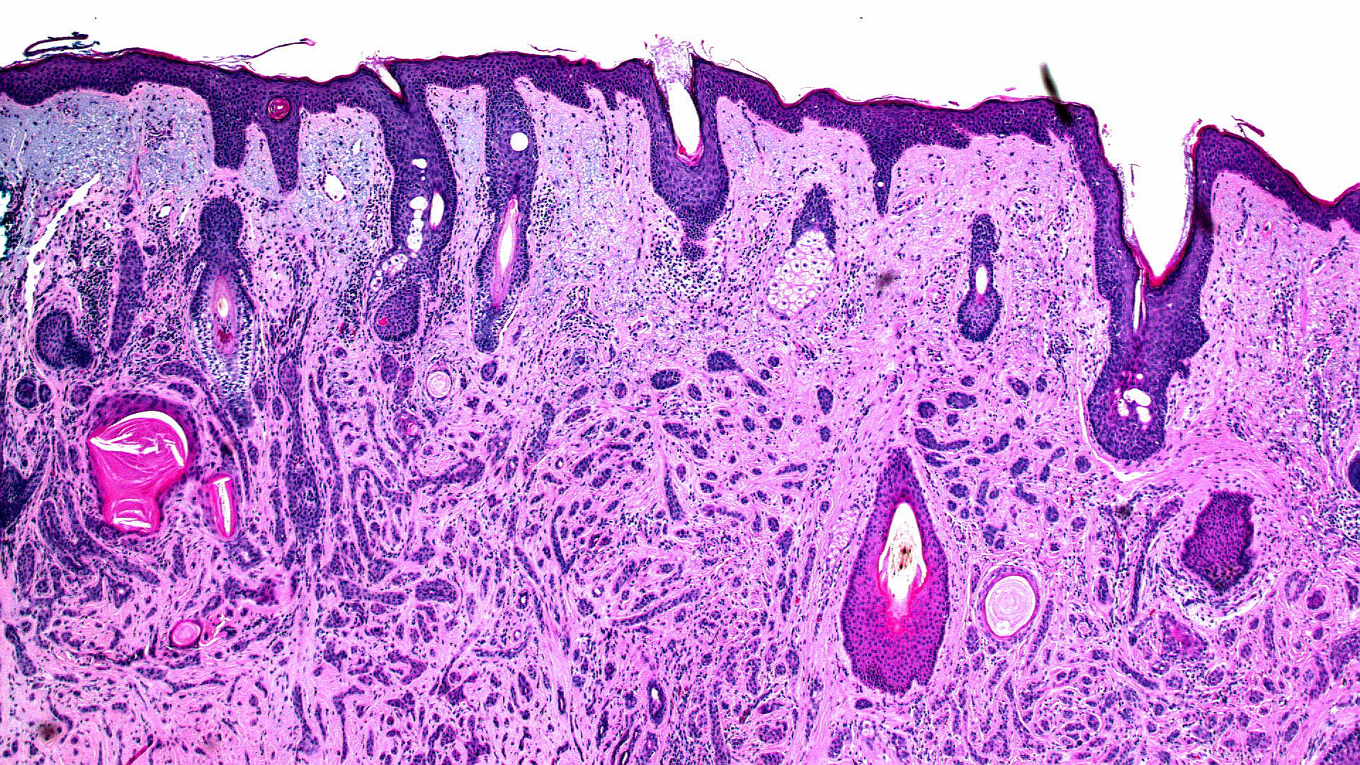
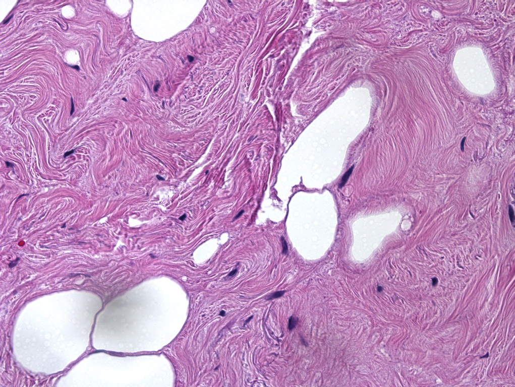
Microcystic Adnexal Carcinoma 40x
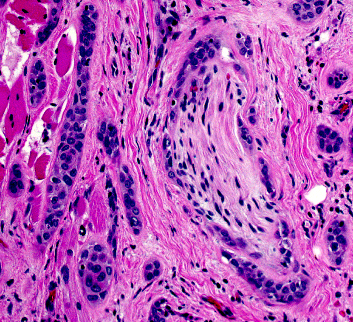
Microcystic Adnexal Carcinoma- Perineural
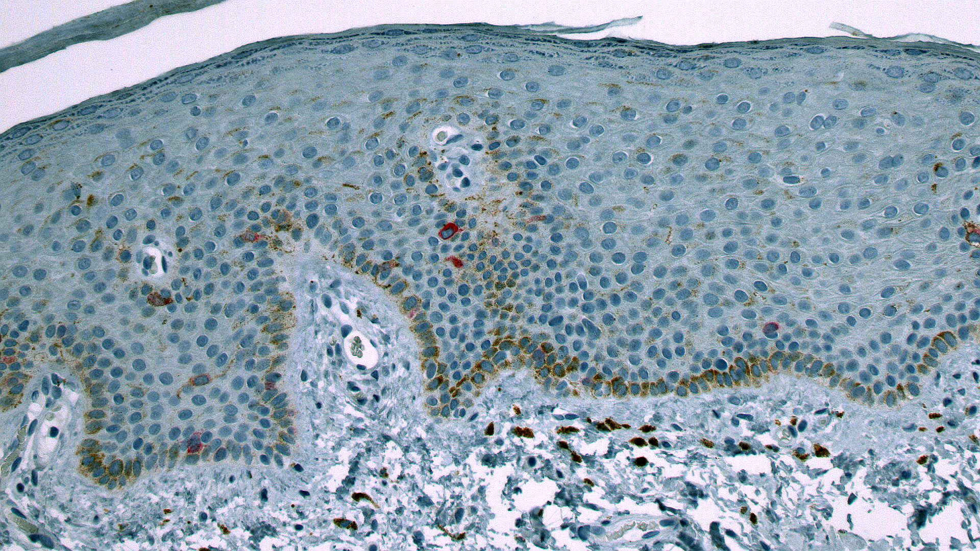
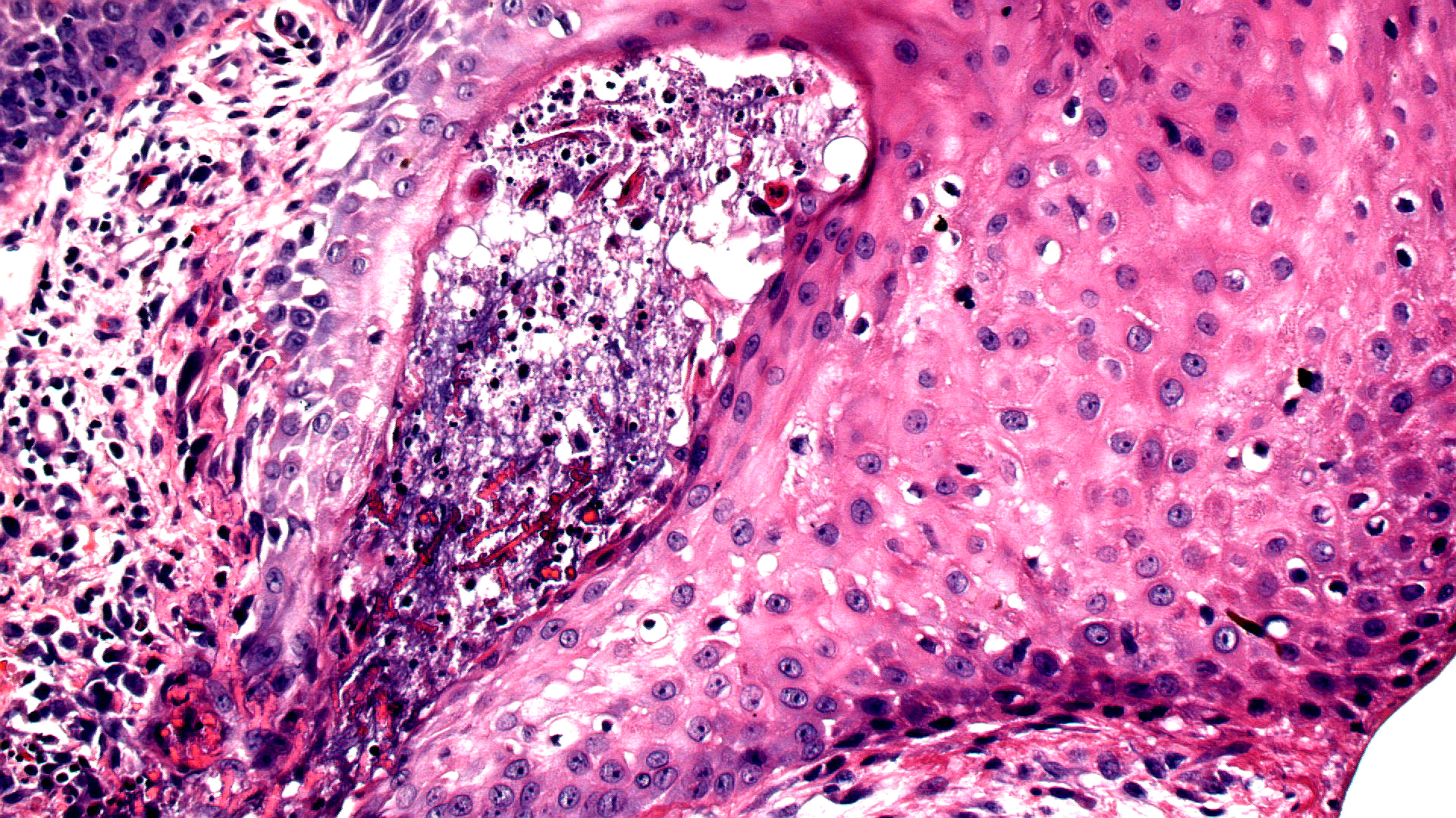
Melanotic Macule Vulva Mart
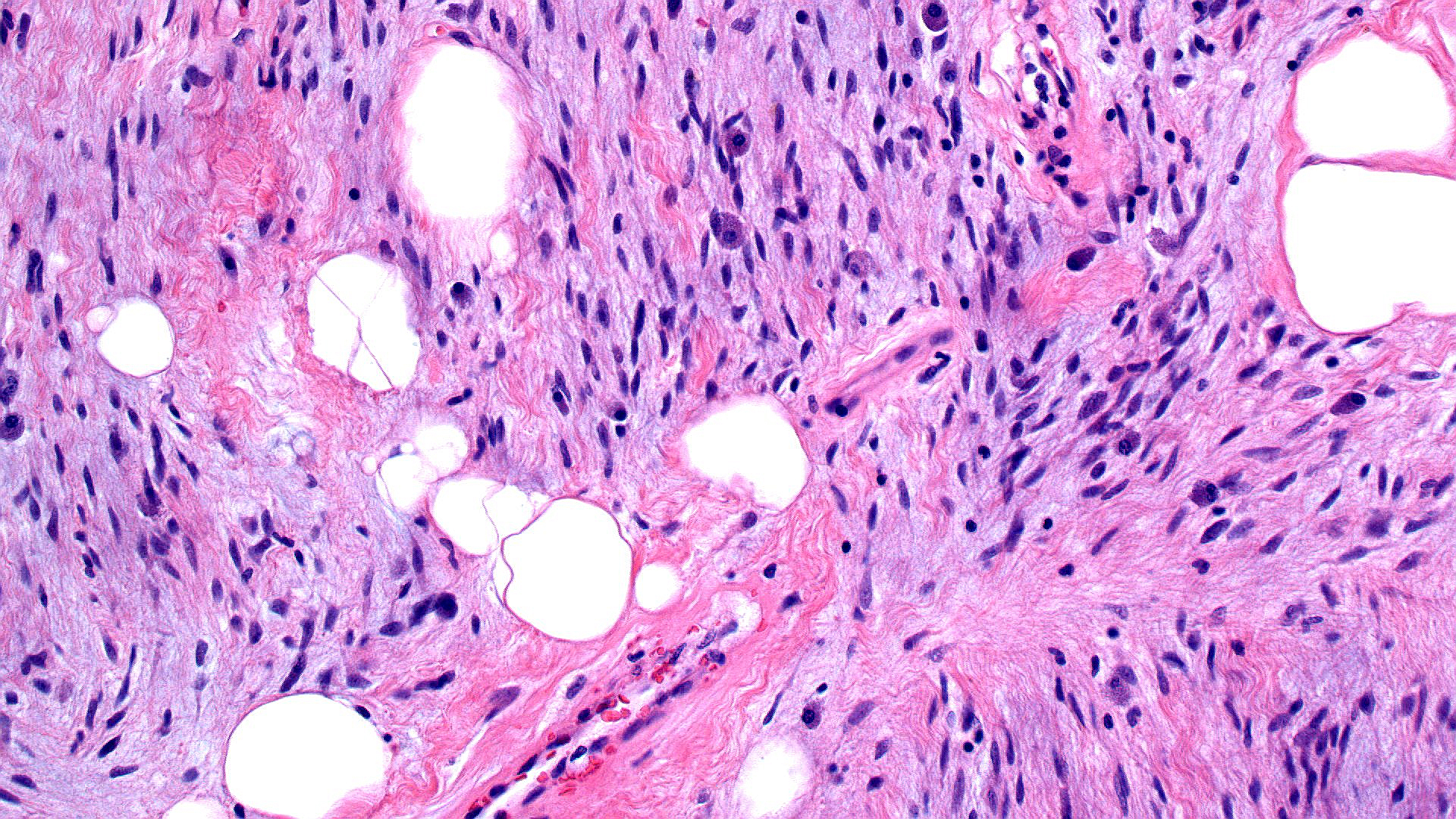
Spindle Cell Lipoma 200x
INFINITY3-1 Sample Images
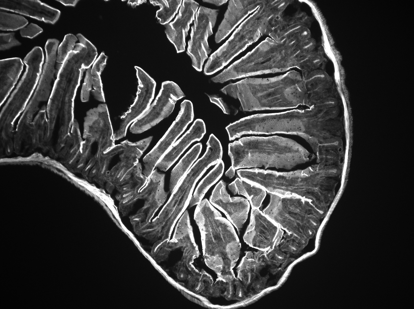
Mouse Intestine in Monochrome
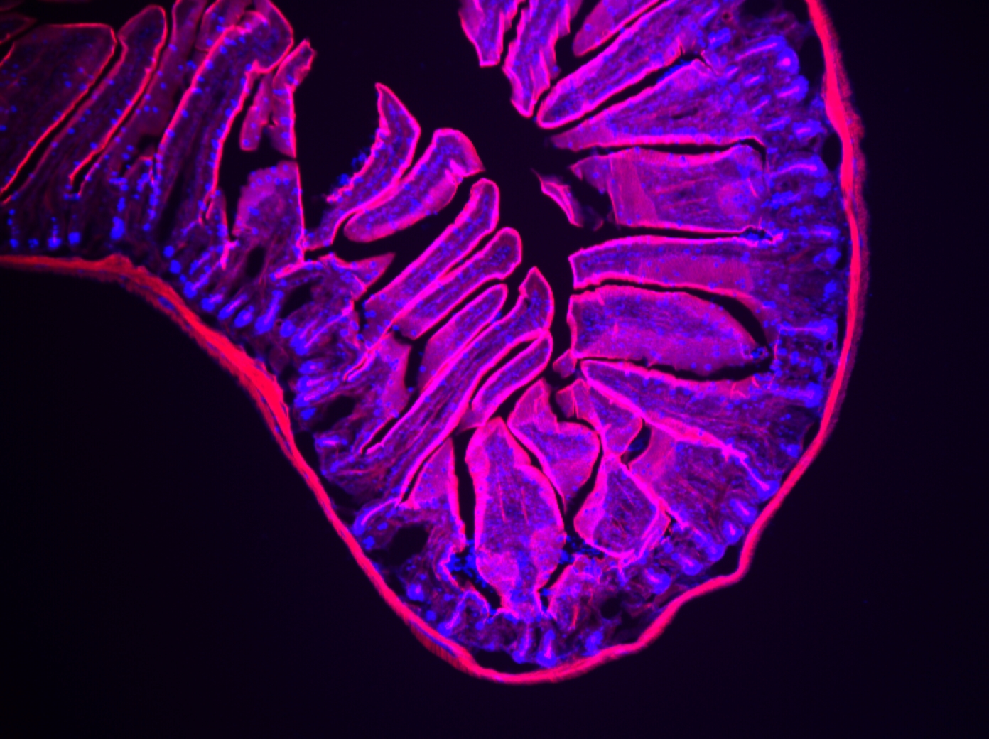
Mouse Intestine Combination
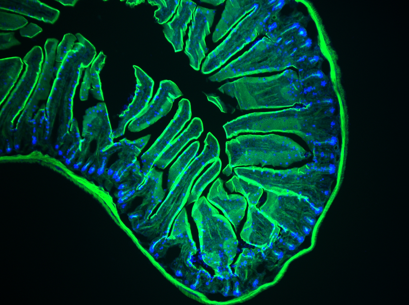
Mouse Intestine Combination

Mouse Endothelioma in Green

Mouse Endothelioma in Green
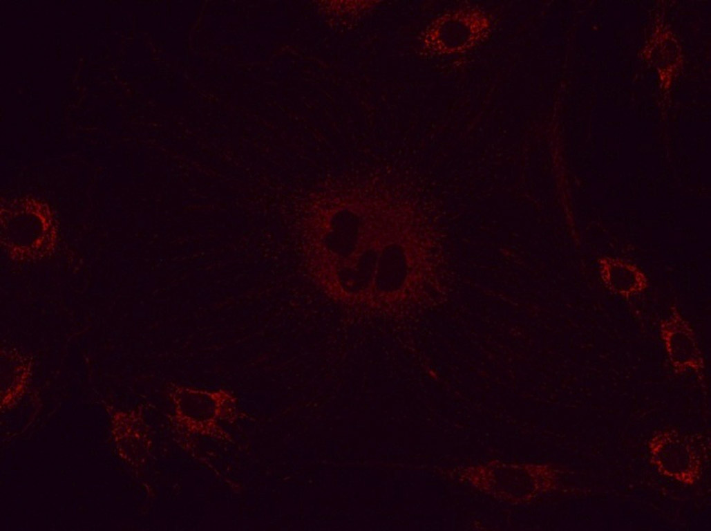
Mouse Endothelioma in Red
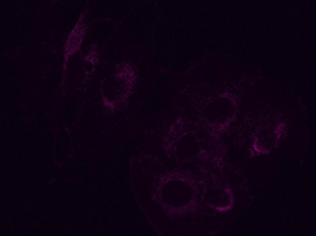
Mouse Endothelioma in Purple
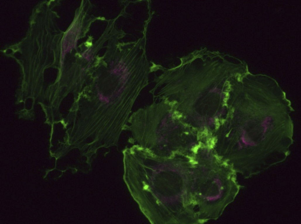
Mouse Endothelioma Merged
Penicillium Mold 4x

Penicillium Mold 10x
Penicillium Mold Vector Merge
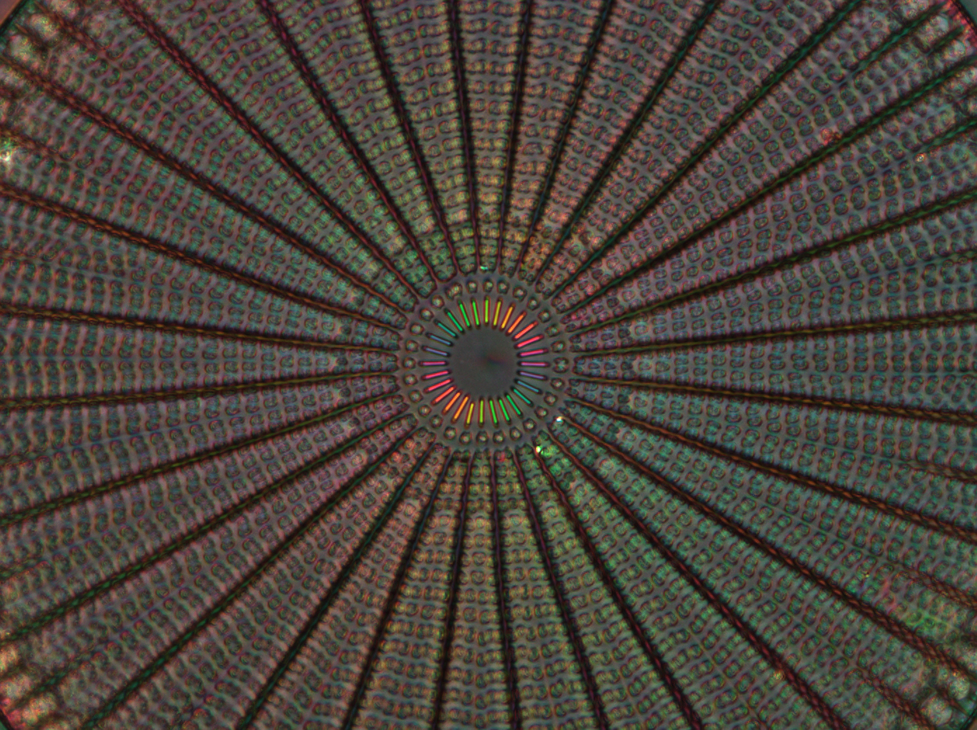
Diatom's Silicified Cell Wall (2015 INFINITY Photo Contest Winner: Image by Michael Shribak, Marine Biological Laboratory)
INFINITY3-1UR Sample Images
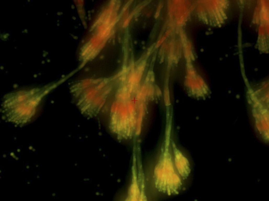
Fluorescence
Fluorescence
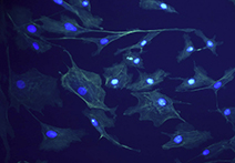
Fluorescence
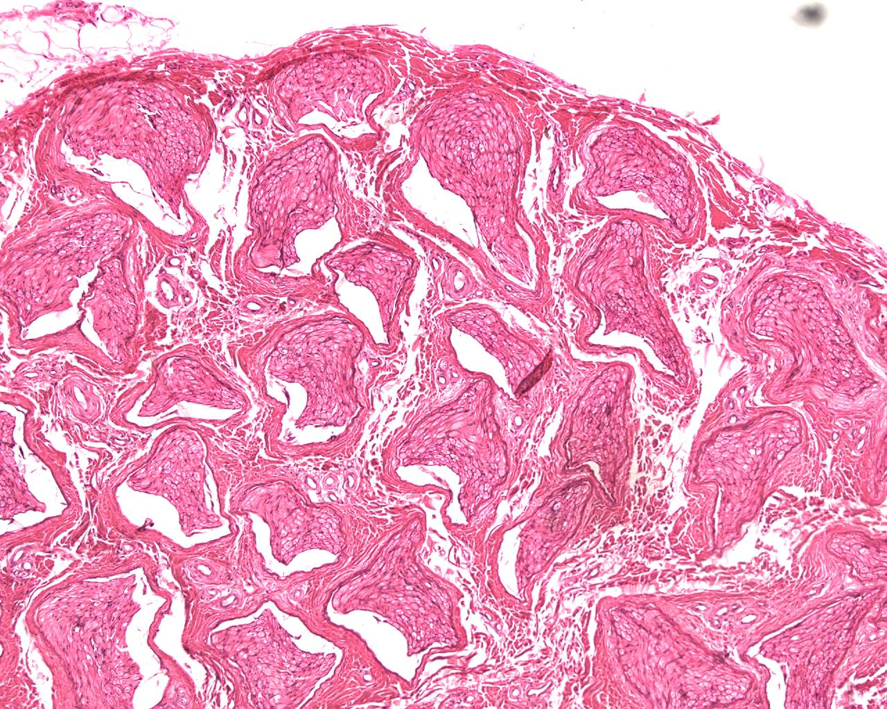
Mammal Peripheral Nerve

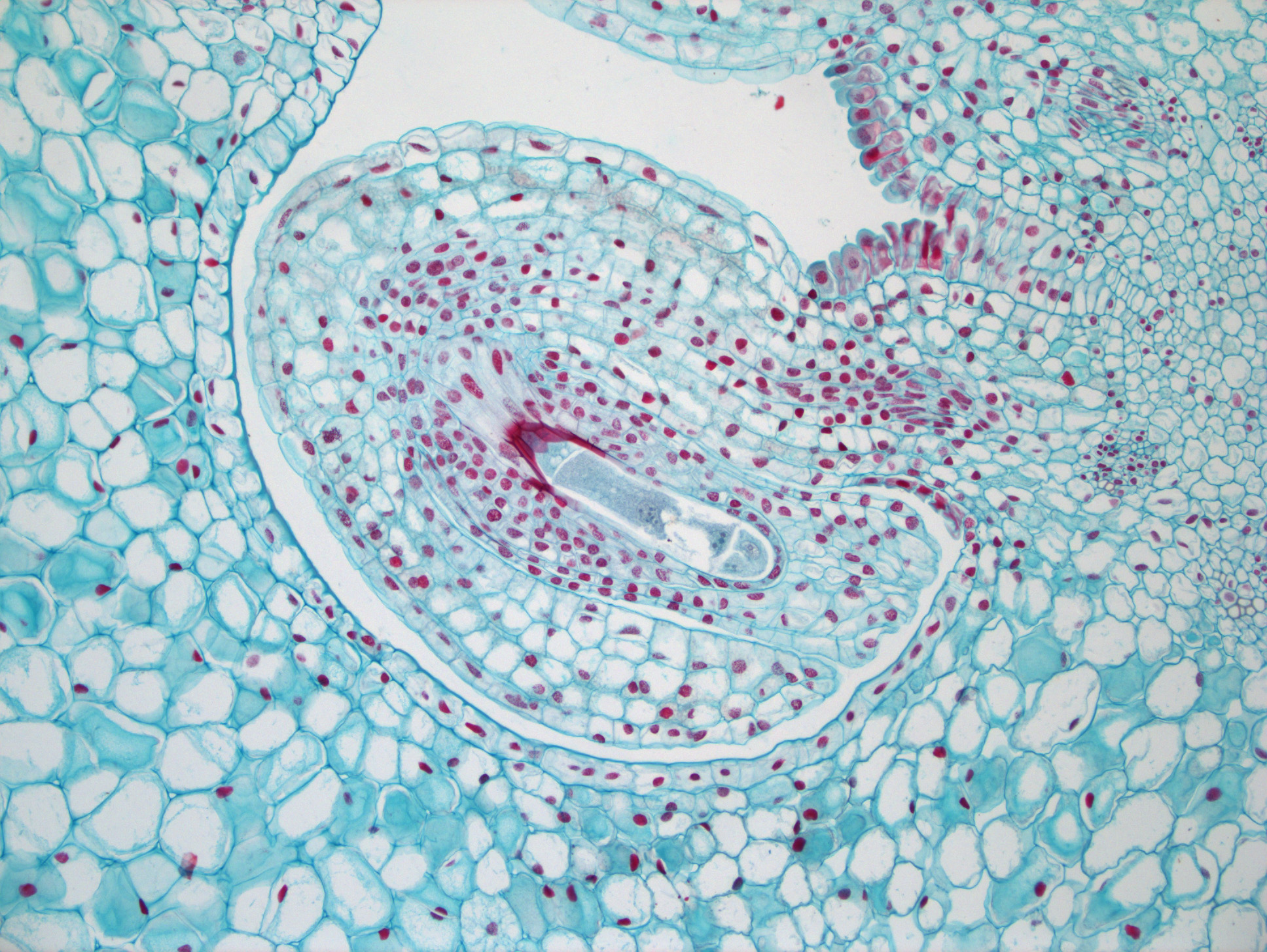
Mammal Ovarian Follicles
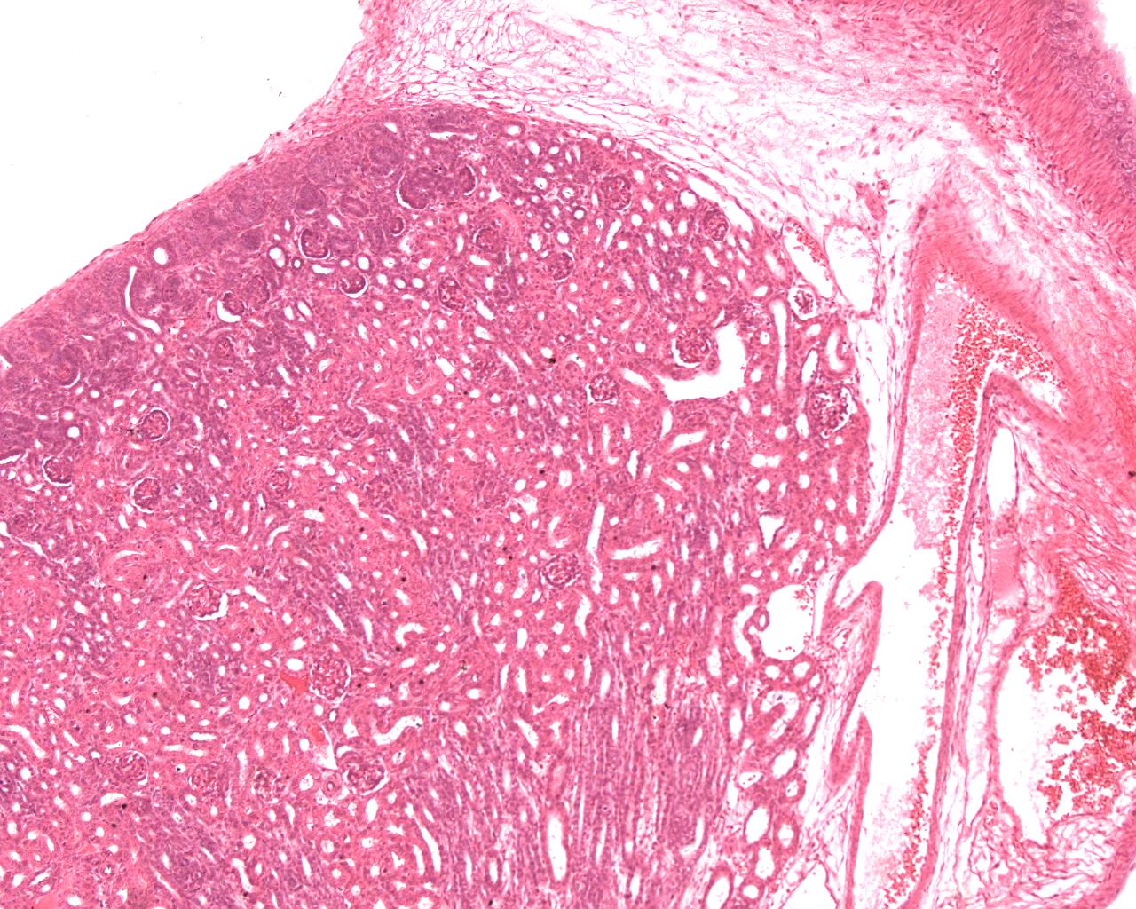
Mammal Kidney
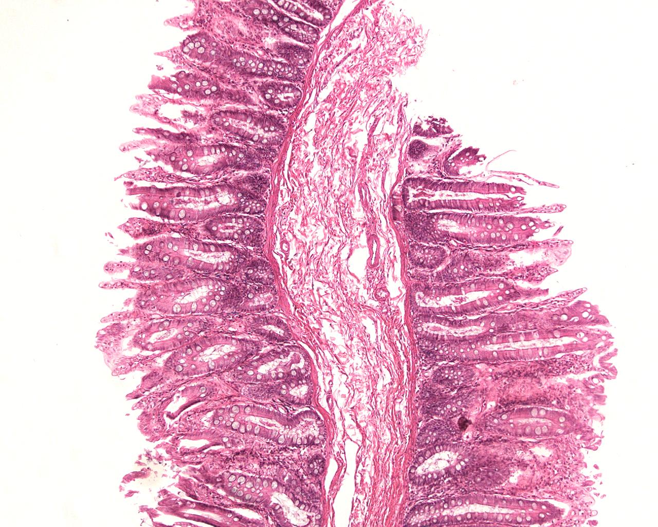
Mammal Colon
INFINITY3-3UR Sample Images
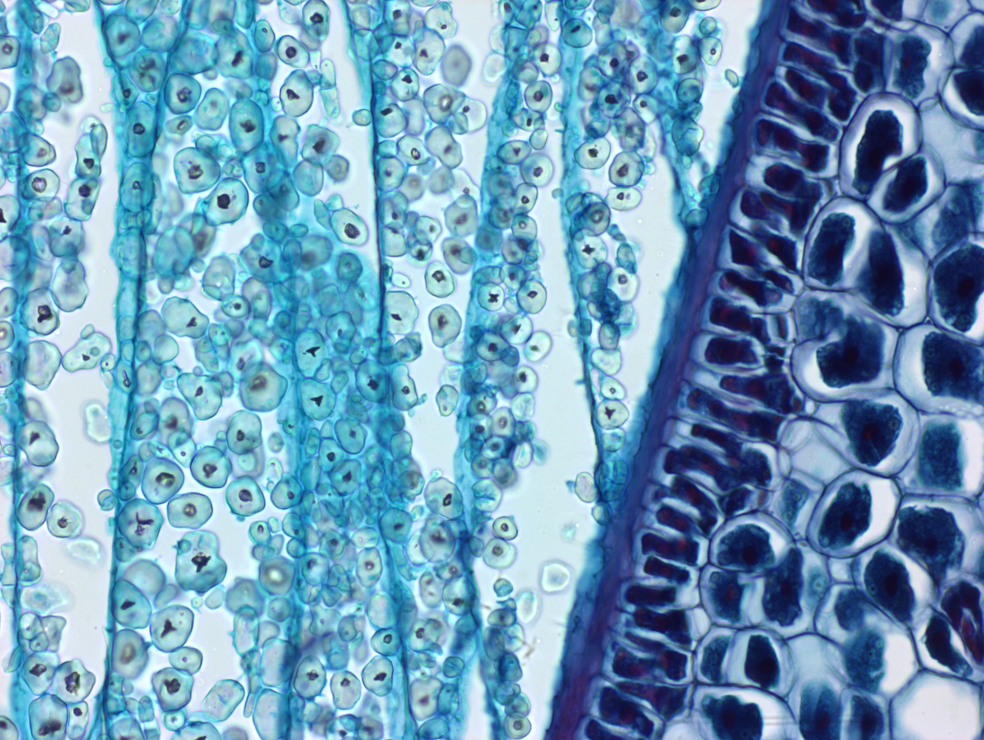
Corn Grain Zea
Corn Grain Zea
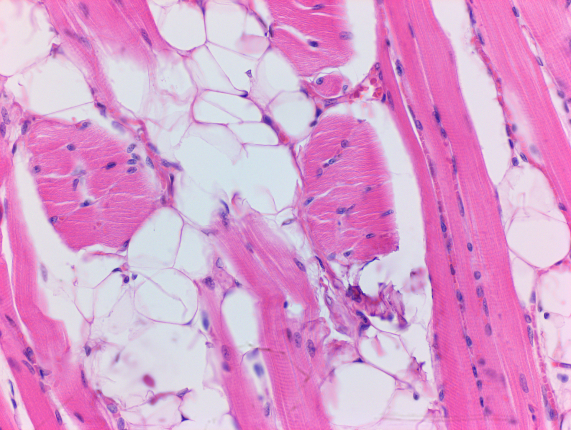
Mammalian Tissue
Ilium Brownii
10x Objective, 10 ms Exposure, 1.2x Gain, 1.0 Gamma

Tissue
40x Objective, 6.75ms Exposure, 1.5x Gain, 1.0 Gamma
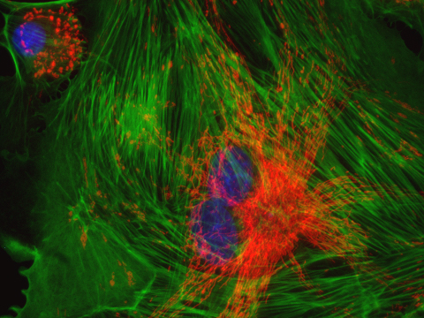
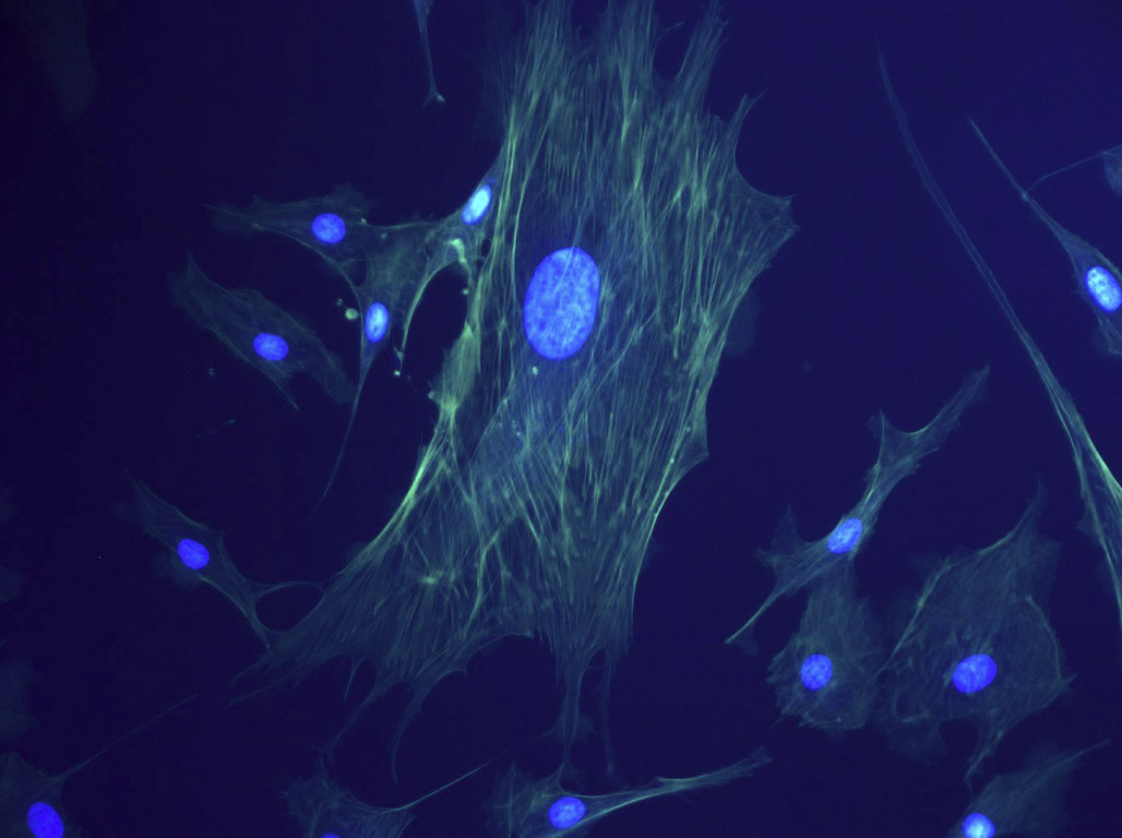
BPAE Fixed Cell
40x Objective, 1.0x Gain
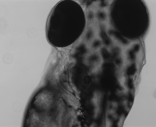
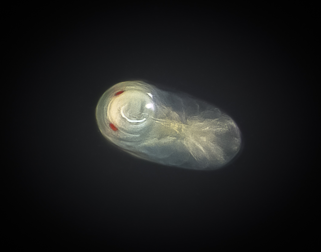
Live Zebra Fish
10x Objective, Brightfield

Parasitoid Wasp (Photo Submission Contest entry by William Fisher)
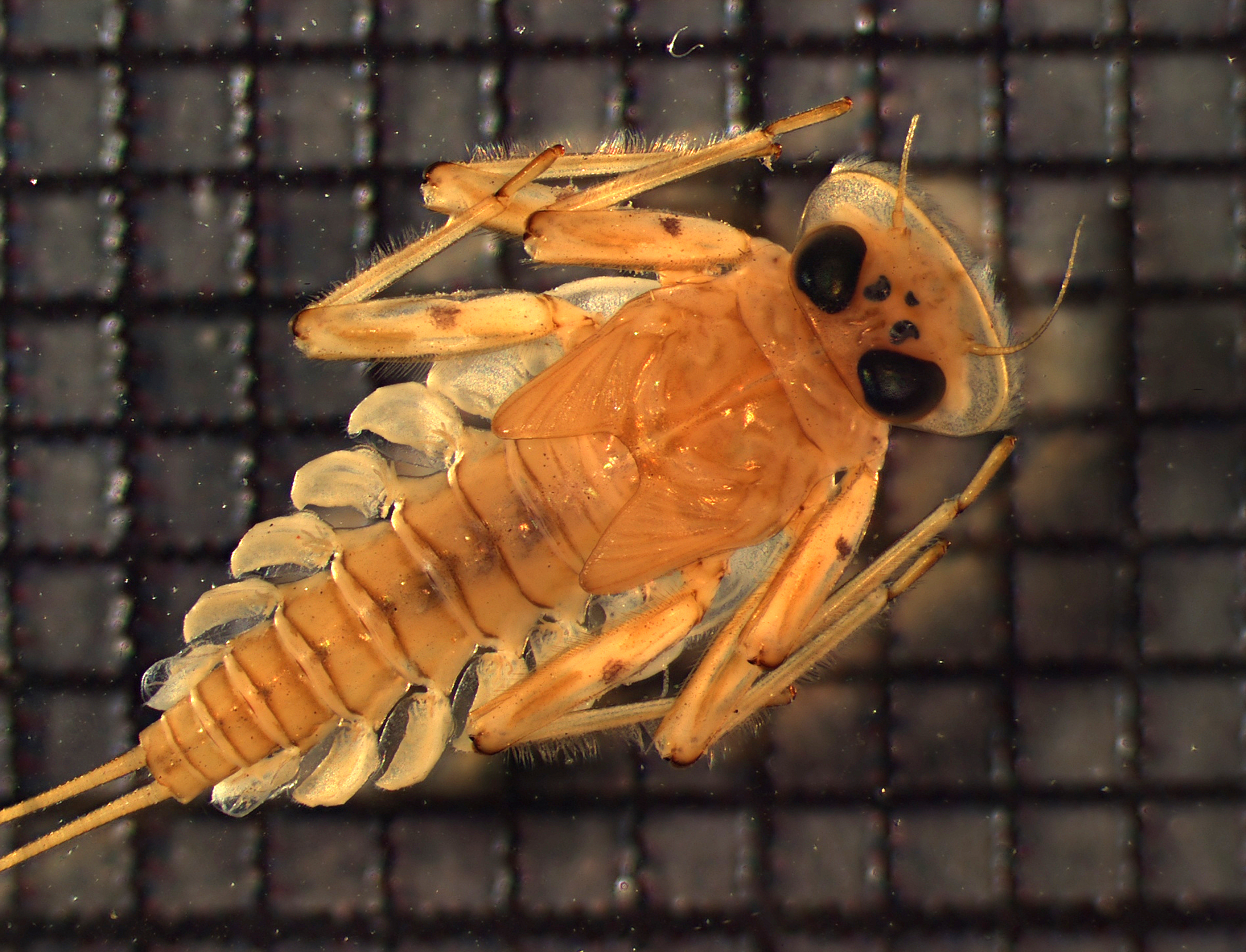
Mayfly (Photo: Morgan Ford)

Diving Beetle (Photo: Morgan Ford)
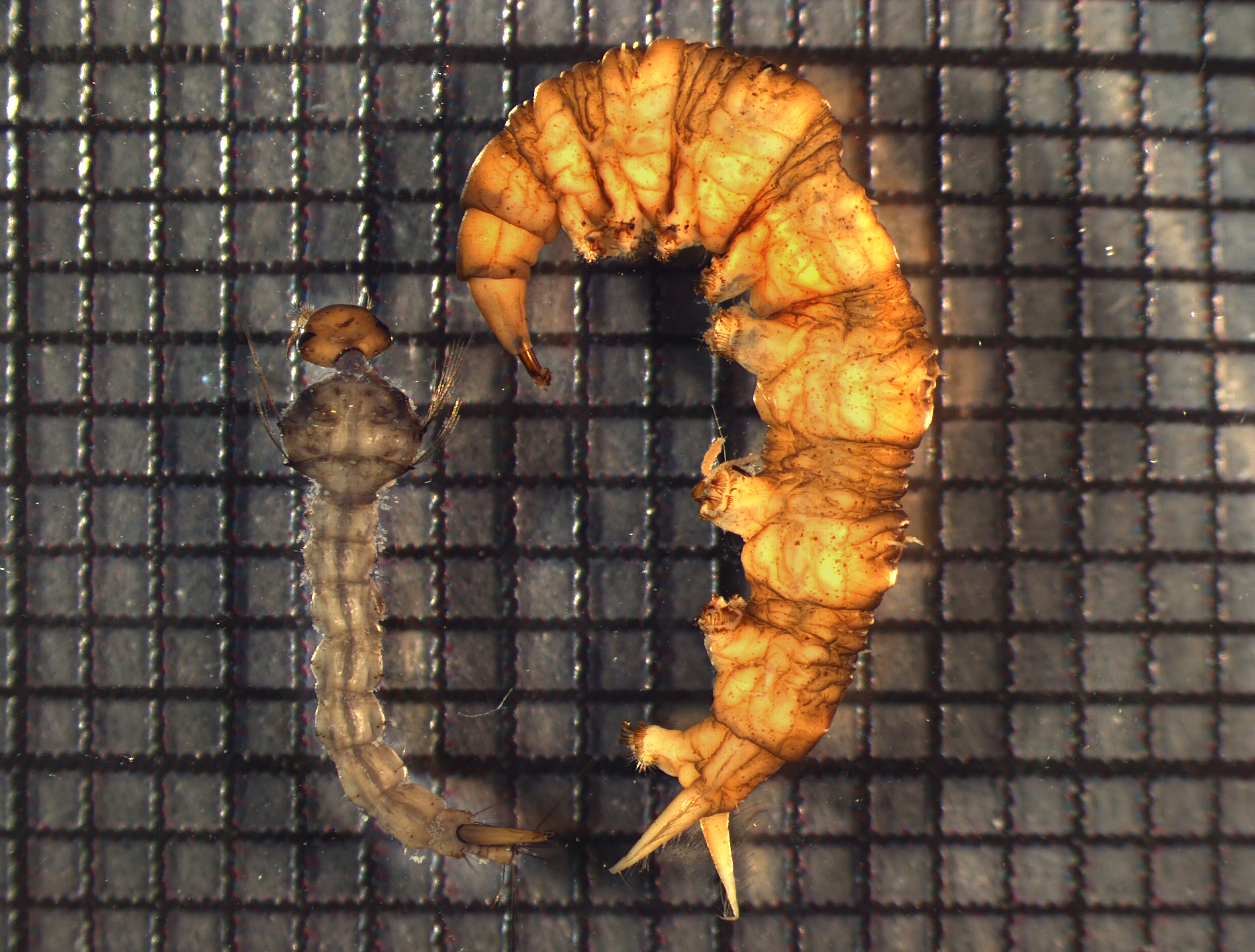
Diptera (Photo: Morgan Ford)
INFINITY3-6UR Sample Images
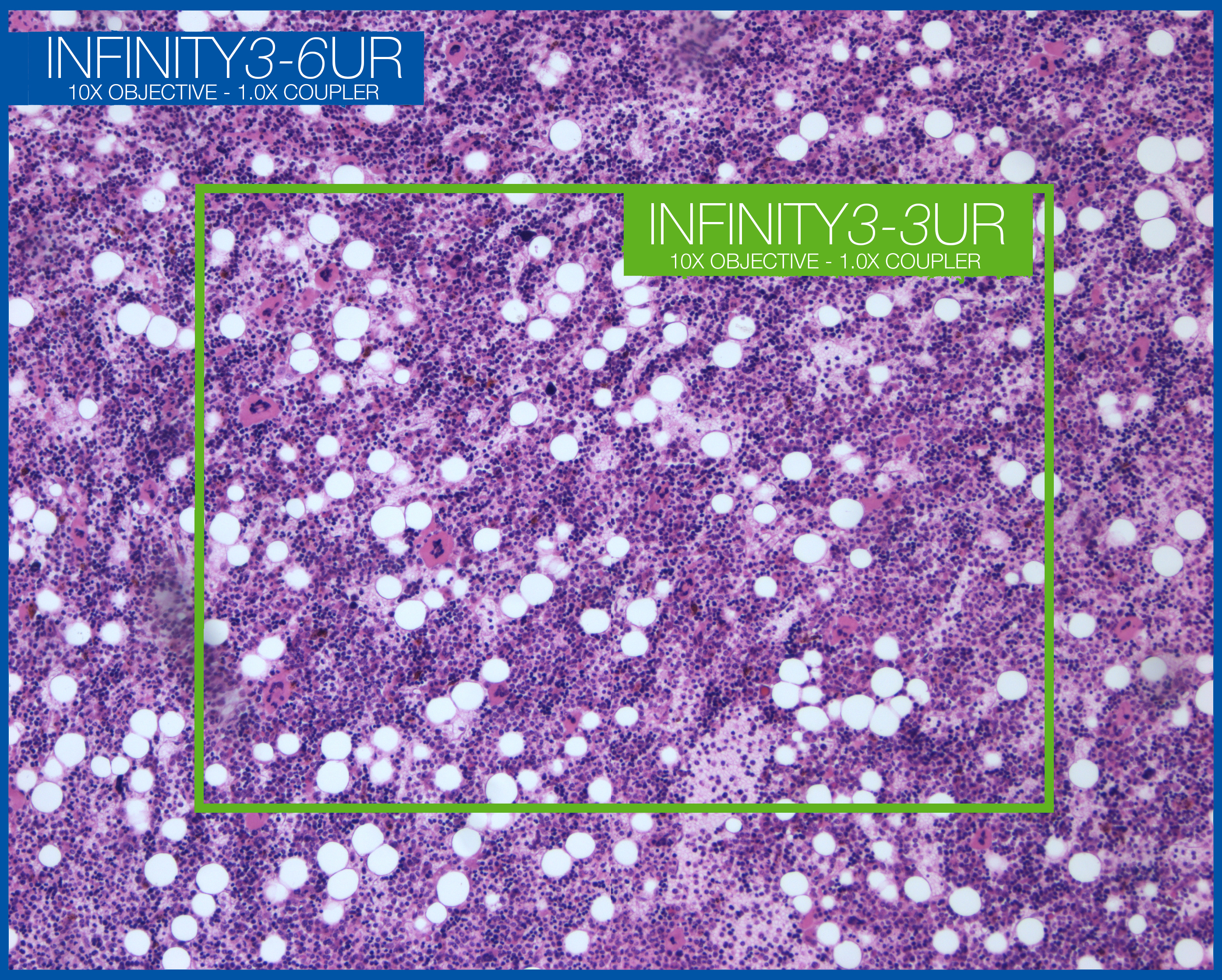
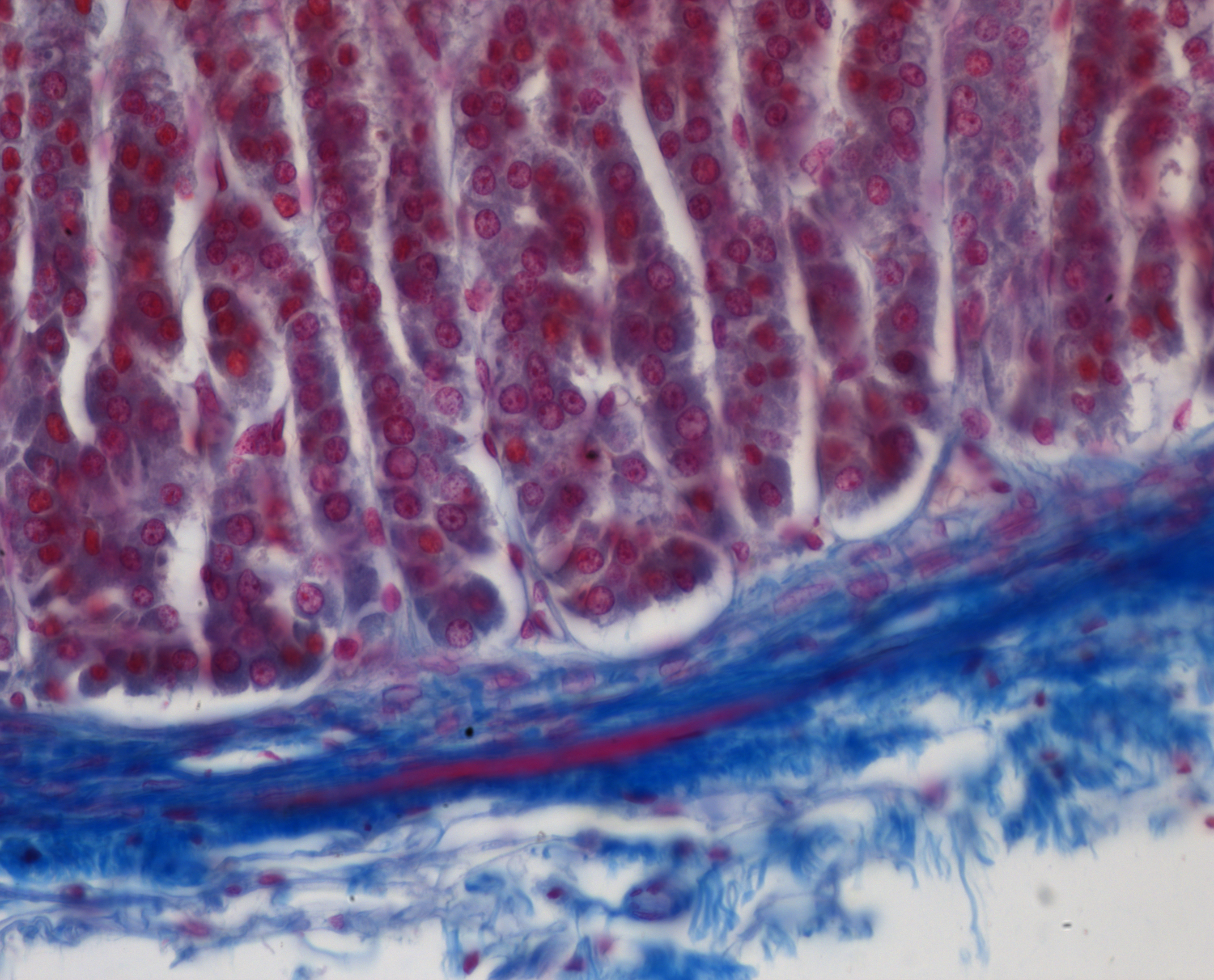
Mammal Bone Marrow with H&E Staining
10x Objective
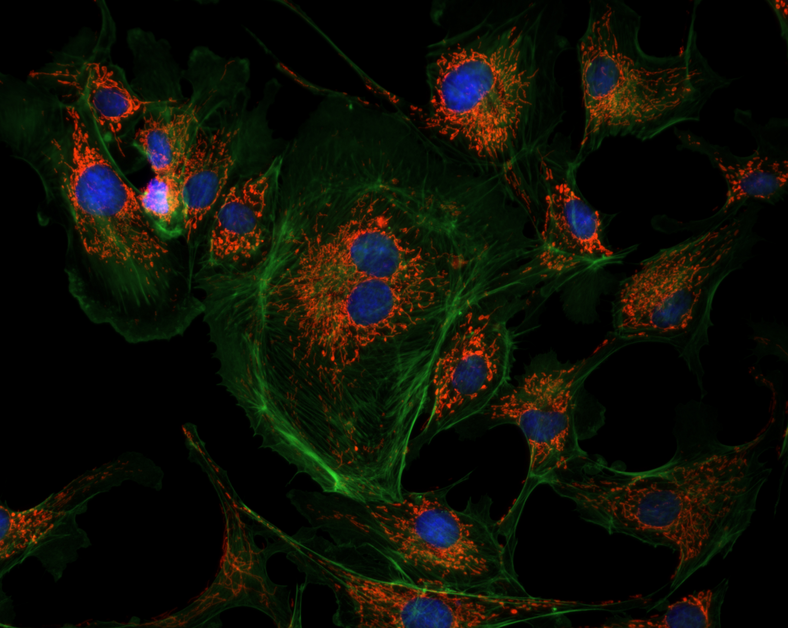
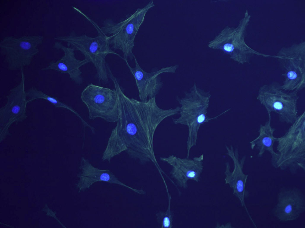
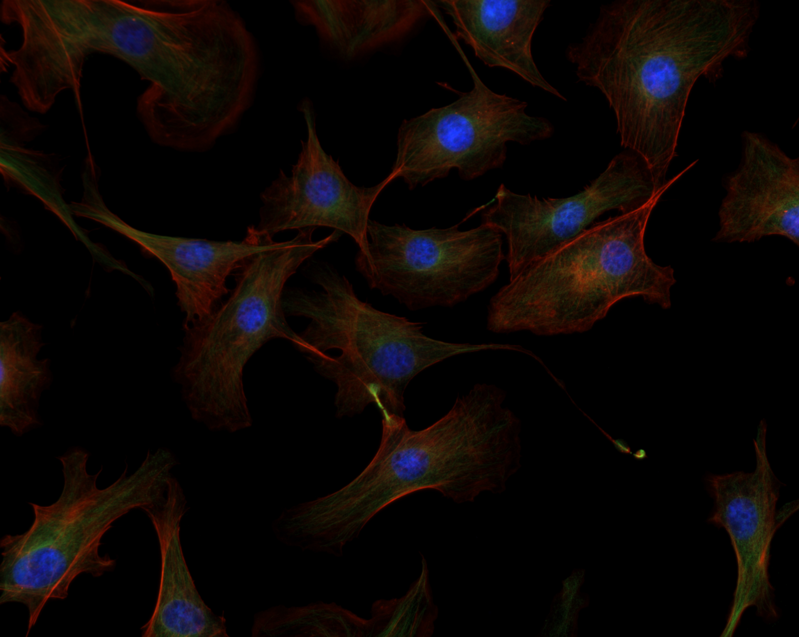
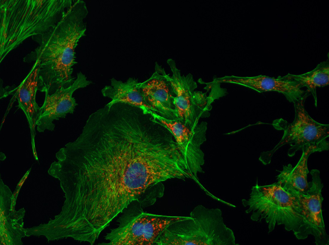
BPAE Sample
40x Objective
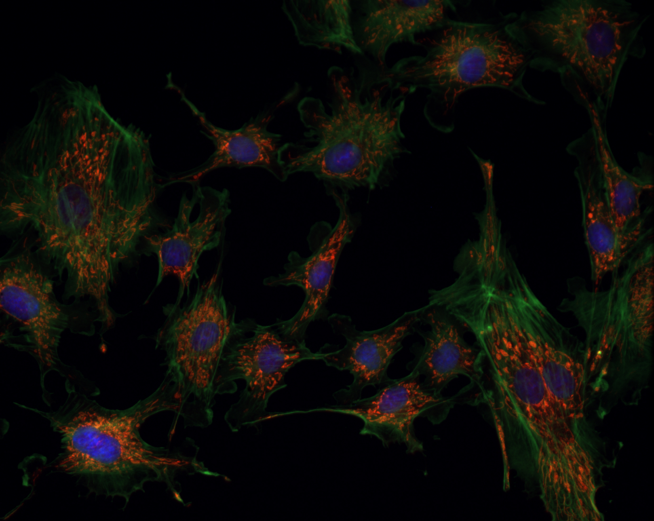
BPAE Sample
40x Objective

Adrenal Gland of a Cat
40x Objective
Adrenal Gland of a Cat
40x Objective
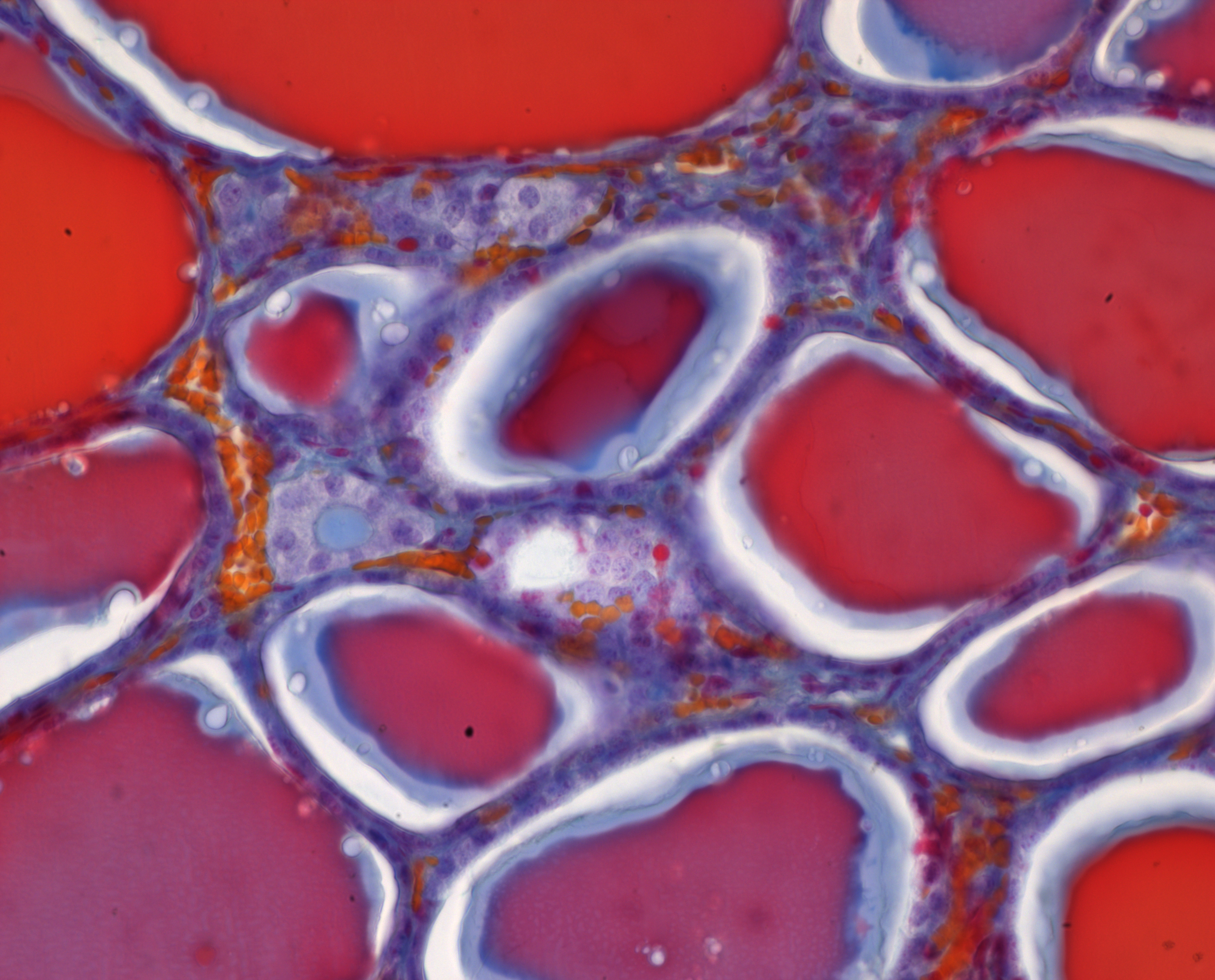
Thyroid Gland of a Sheep
40x Objective
Mouse Kidney
40x Objective
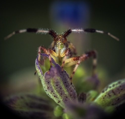
Unknown Hemipteran on Bog Sage (Photo Submission Contest entry by William Fisher)
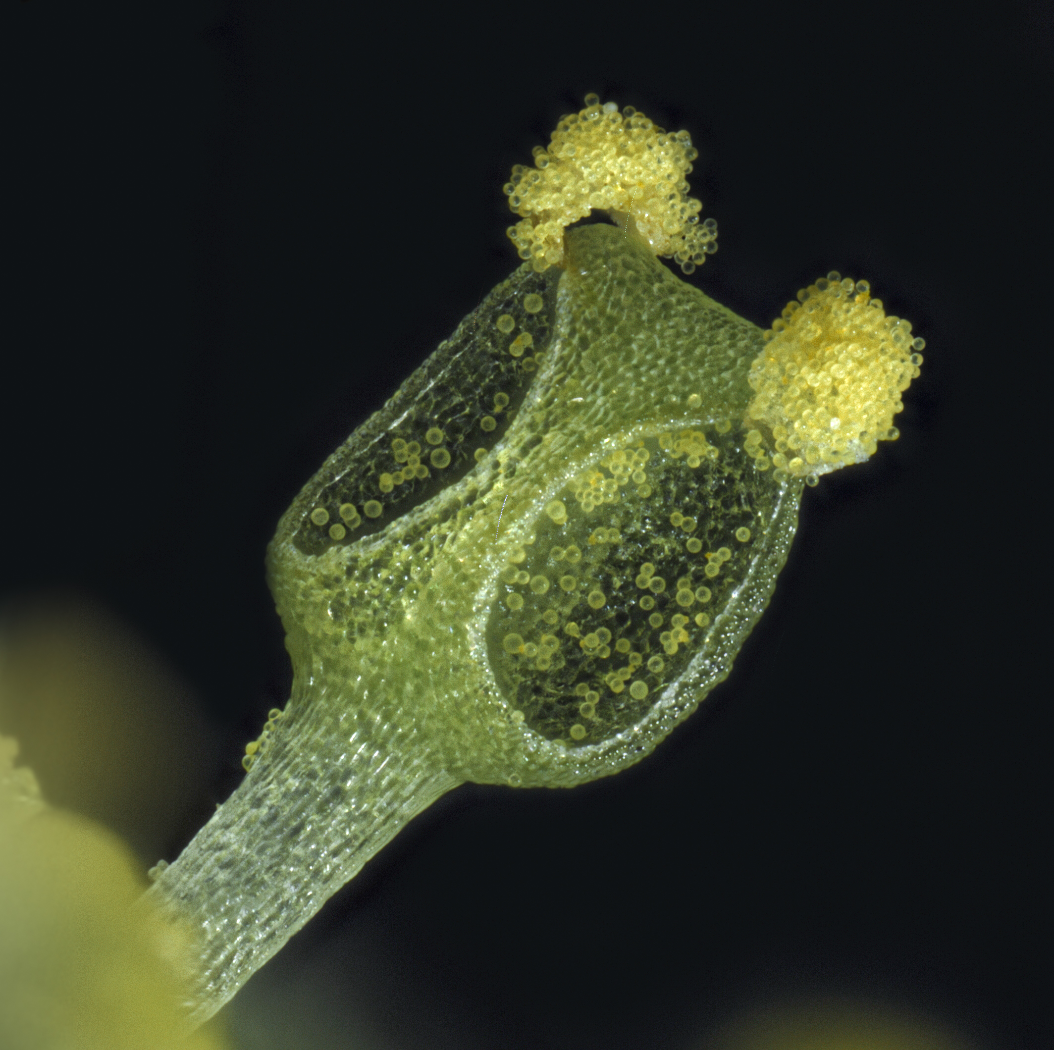
Pollen structure from a flower of the Spicebush (Lindera benzoin), (Photo by William Fisher)
INFINITY3S-1UR Sample Images
BPAE sample
40x Objective
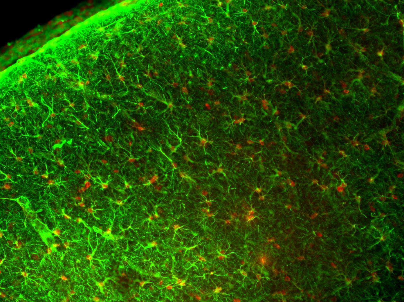
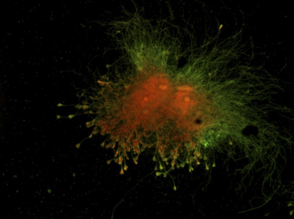
Composite image of the brain of a rat
10x Objective
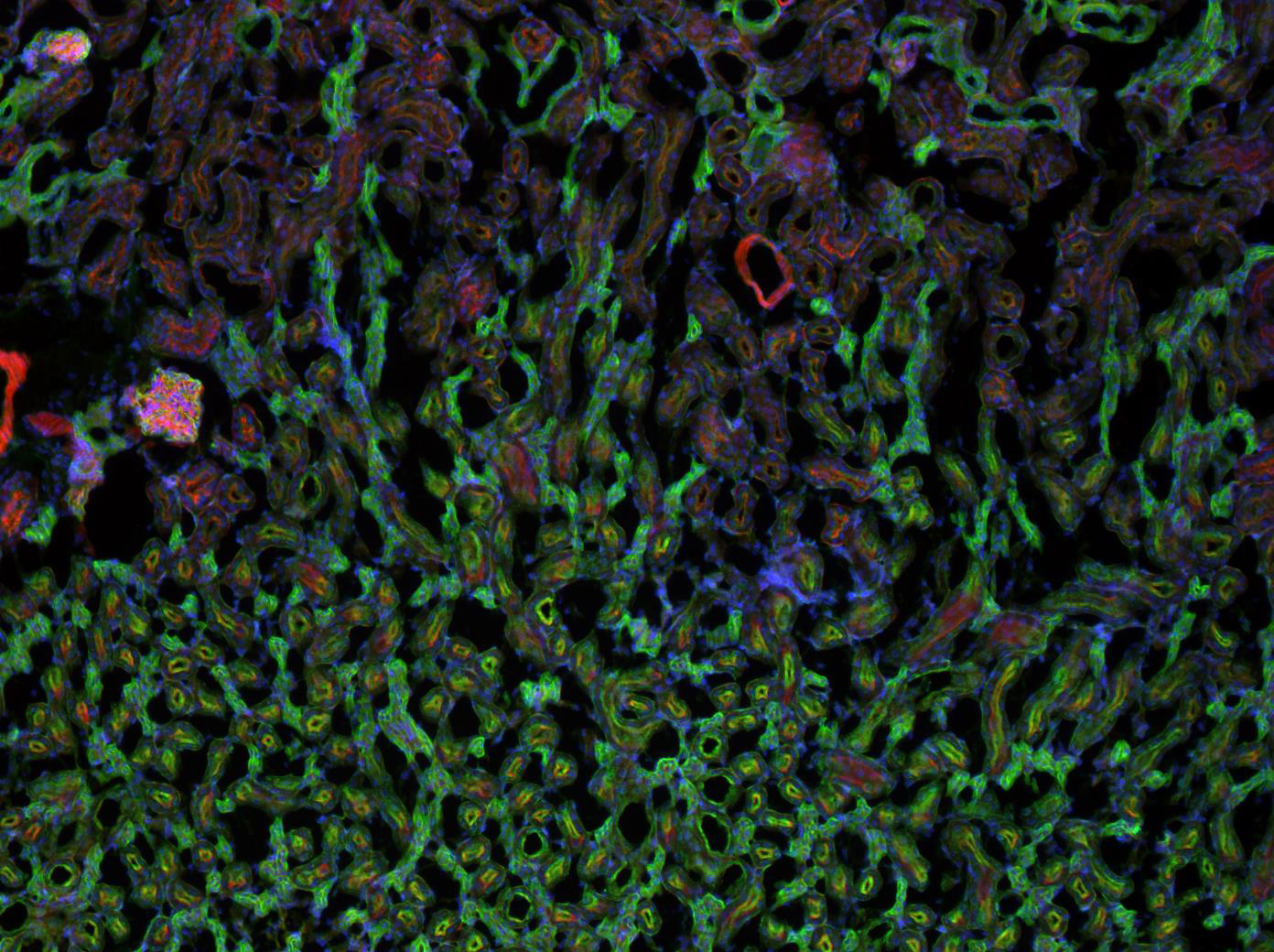
Composite image of a mouse kidney
10x Objective
INFINITY4-11 Sample Images

Tilia
INFINITYX-21 Sample Images*
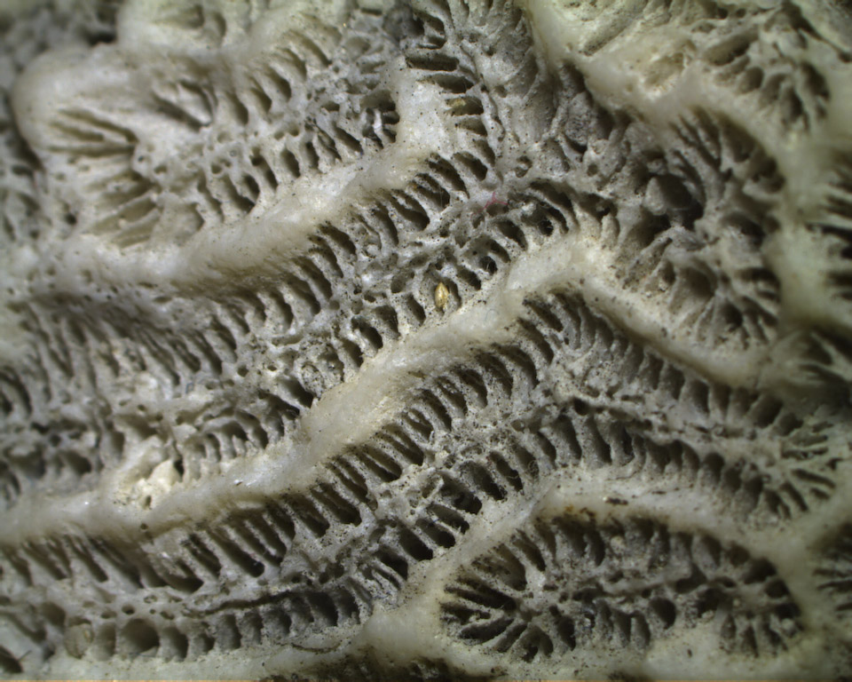
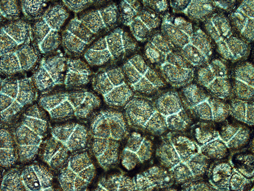
Coral

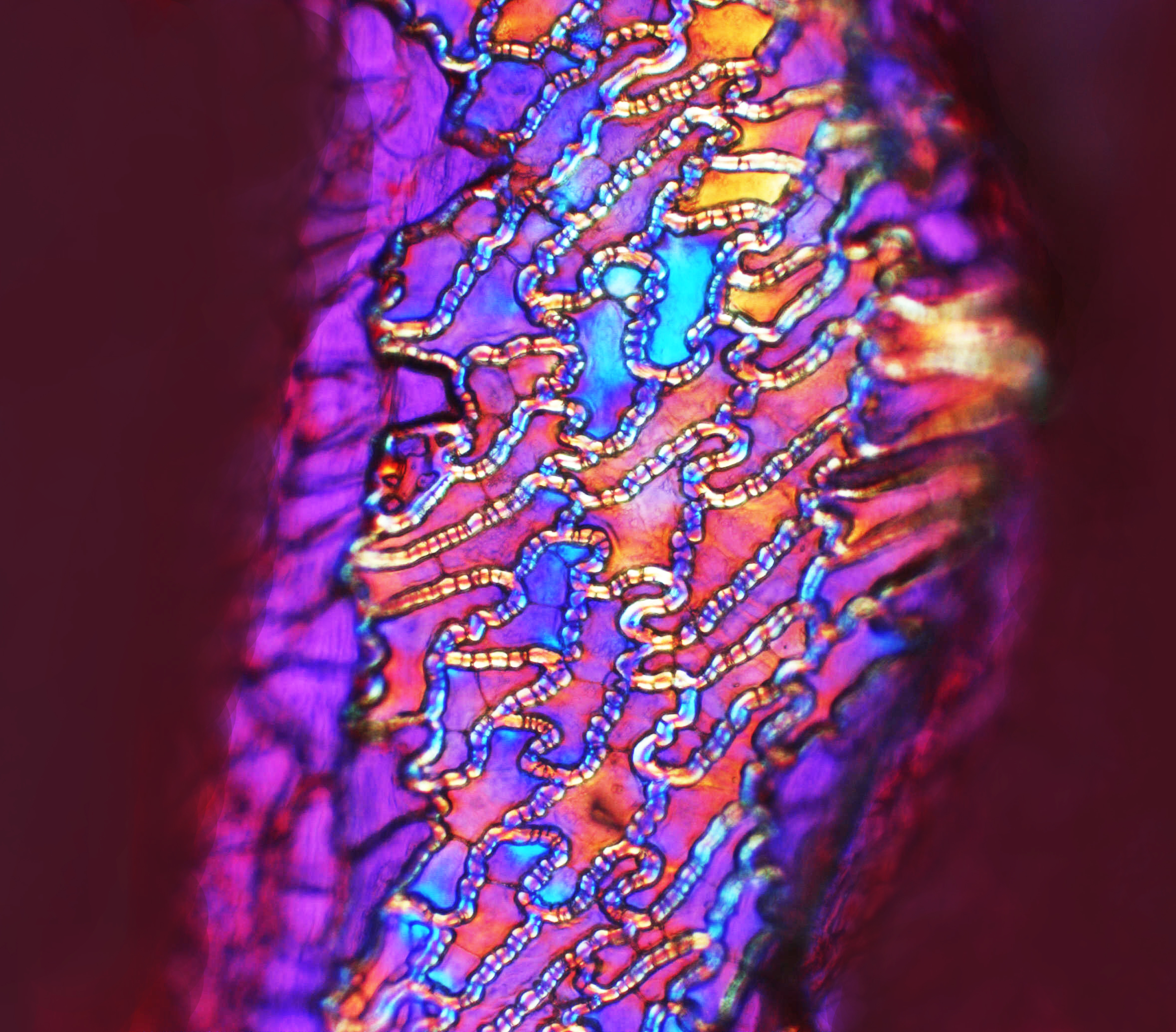
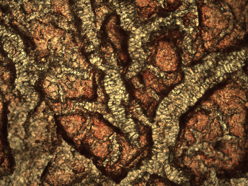
Fosil
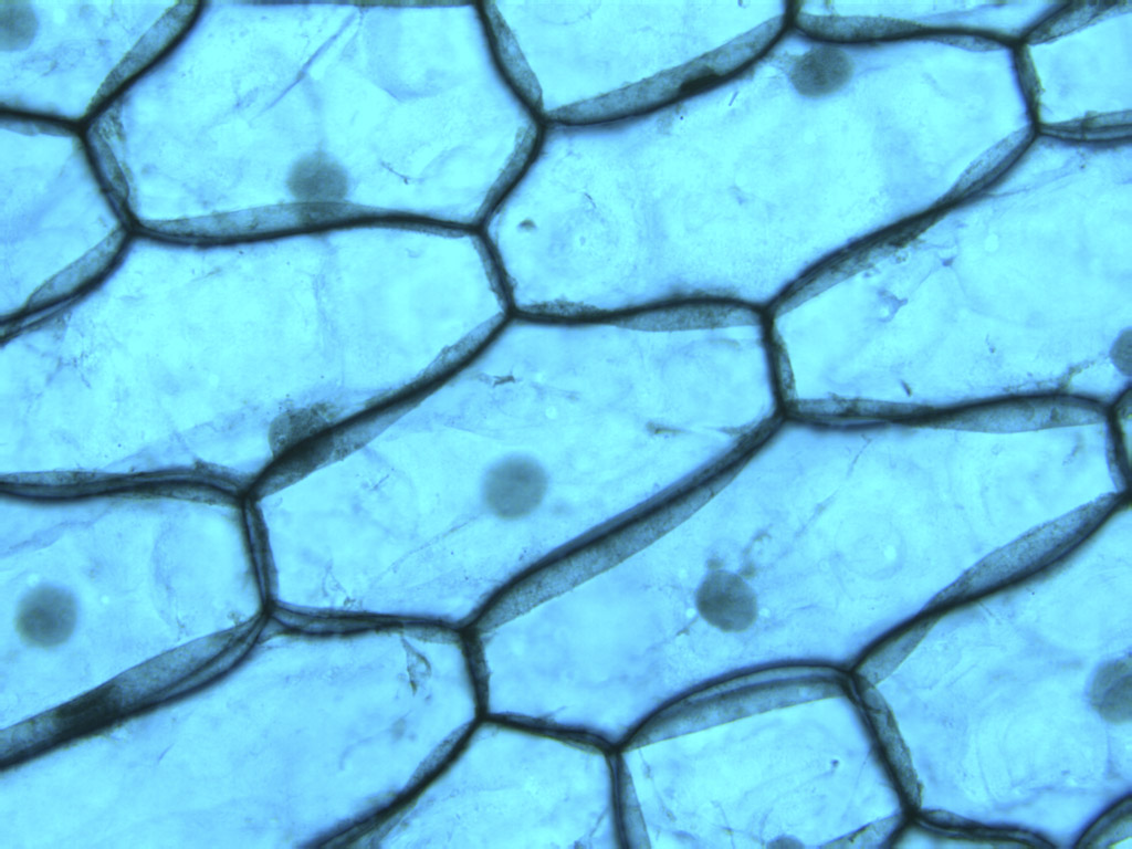
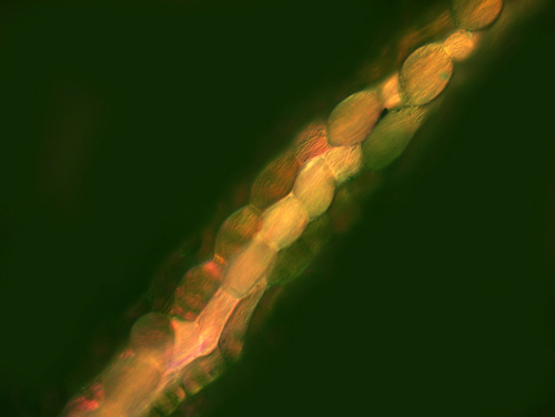
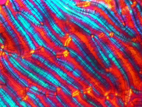
Leaf
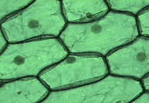
Leaf
* Please note this camera is no longer a part of our portfolio.
INFINITYX-32 Sample Images
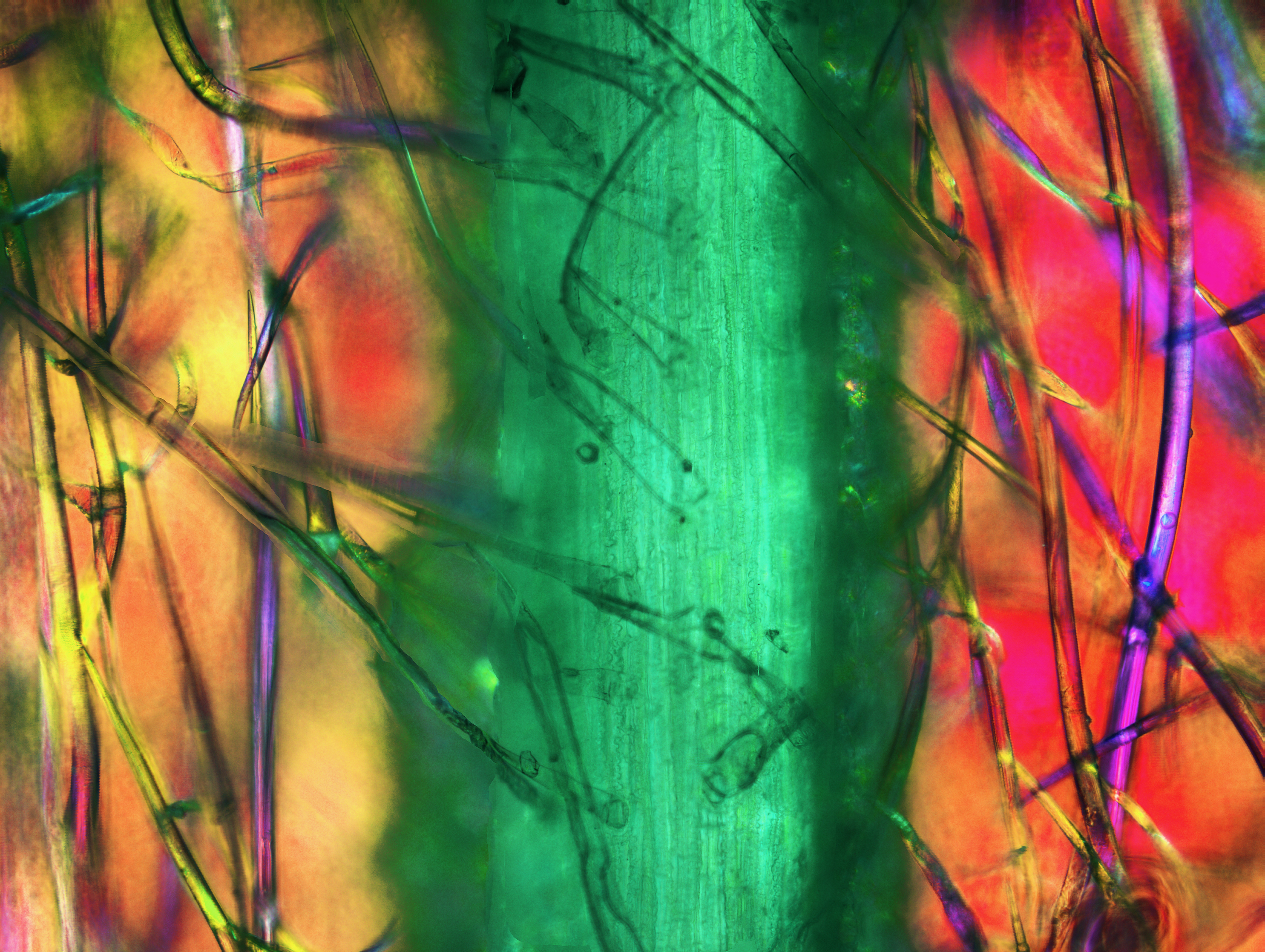
Red Pepper Endocarp (Photo by Robert Rock Belliveau)
Skin of a Green Olive (Photo by Robert Rock Belliveau)
Vein of a Parsley Leaf (Photo by Robert Rock Belliveau)
Blueberry (Photo by Robert Rock Belliveau)
Pear Skin (Photo by Robert Rock Belliveau)

Red Grape (Photo by Robert Rock Belliveau)
Corn Husk with Corn Silk (Photo by Robert Rock Belliveau)

Surface of the Seed of the Common Tomato (Photo by Robert Rock Belliveau)
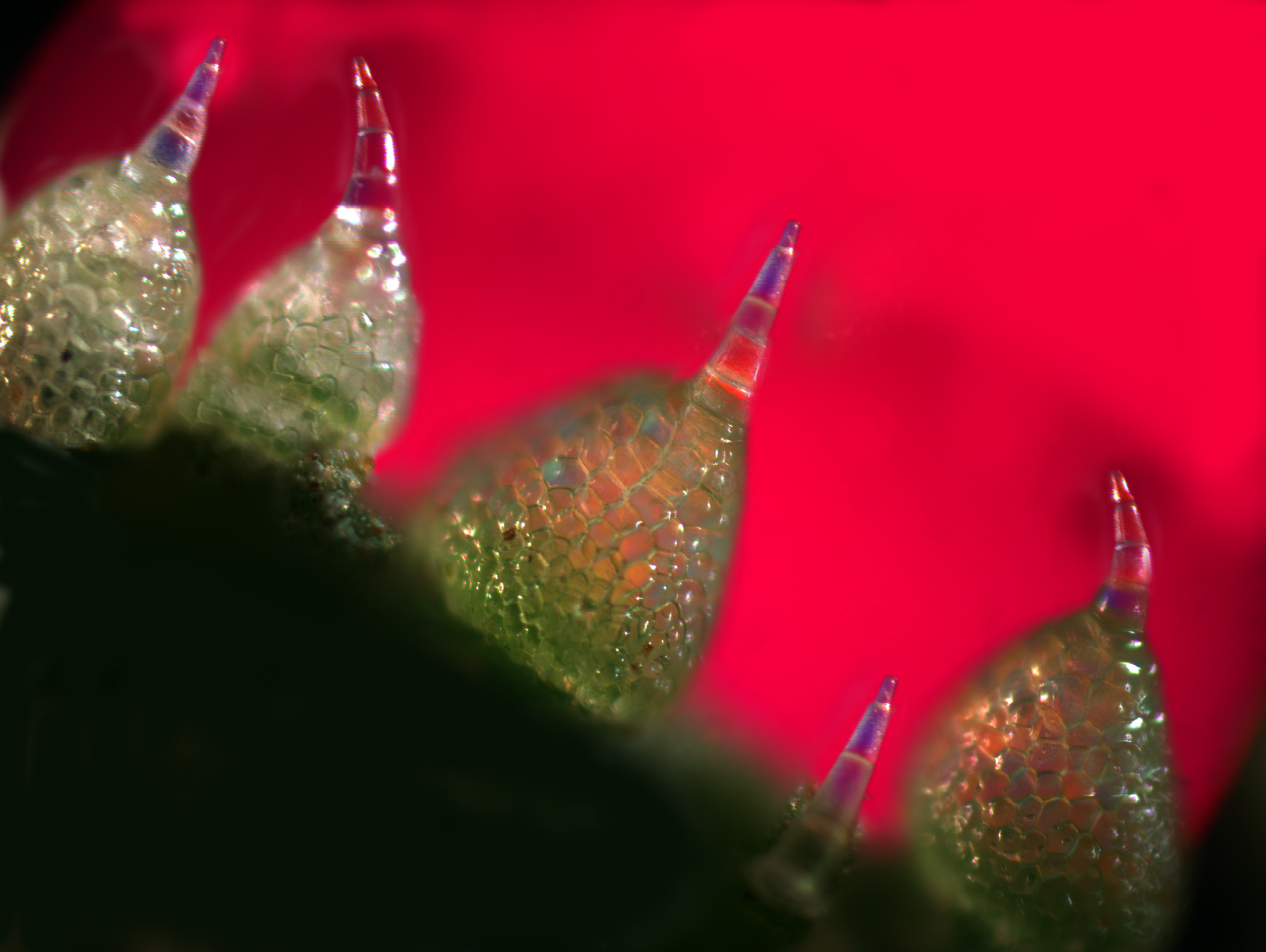
Cucumber Skin with Trichomes (Photo by Robert Rock Belliveau)
Stained Tissue
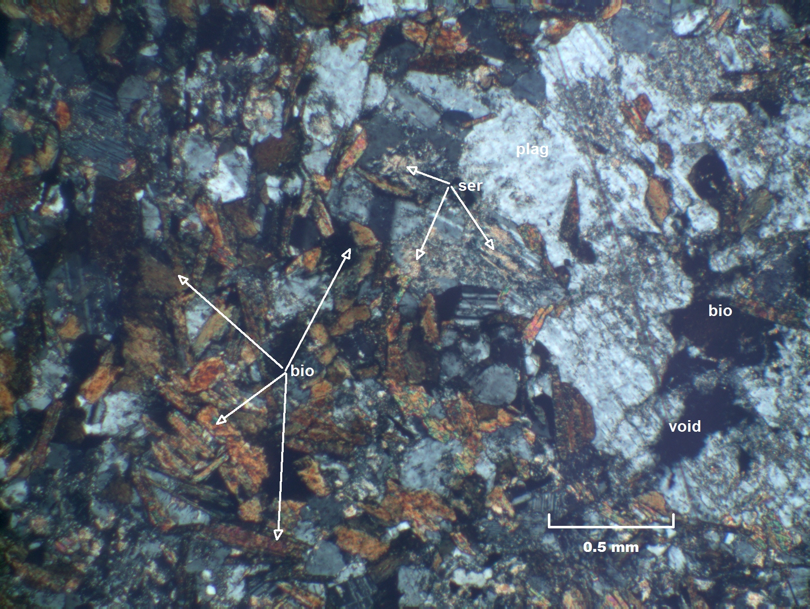
Biotite, Plagioclase, Gneiss
4x Magnification, Crossed polarizers, 5ms Exposure, 2x Gain, 2.3 Gamma (Photo: Terry Crump)
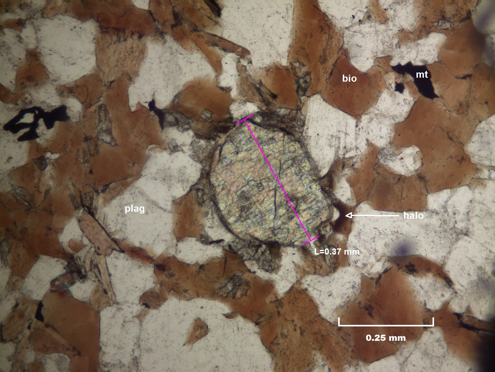
Zircon Crystal in Gneiss
10x Magnification, Plain polarized light, 1.2ms Exposure, 2x Gain, 1.25 Gamma (Photo: Terry Crump)
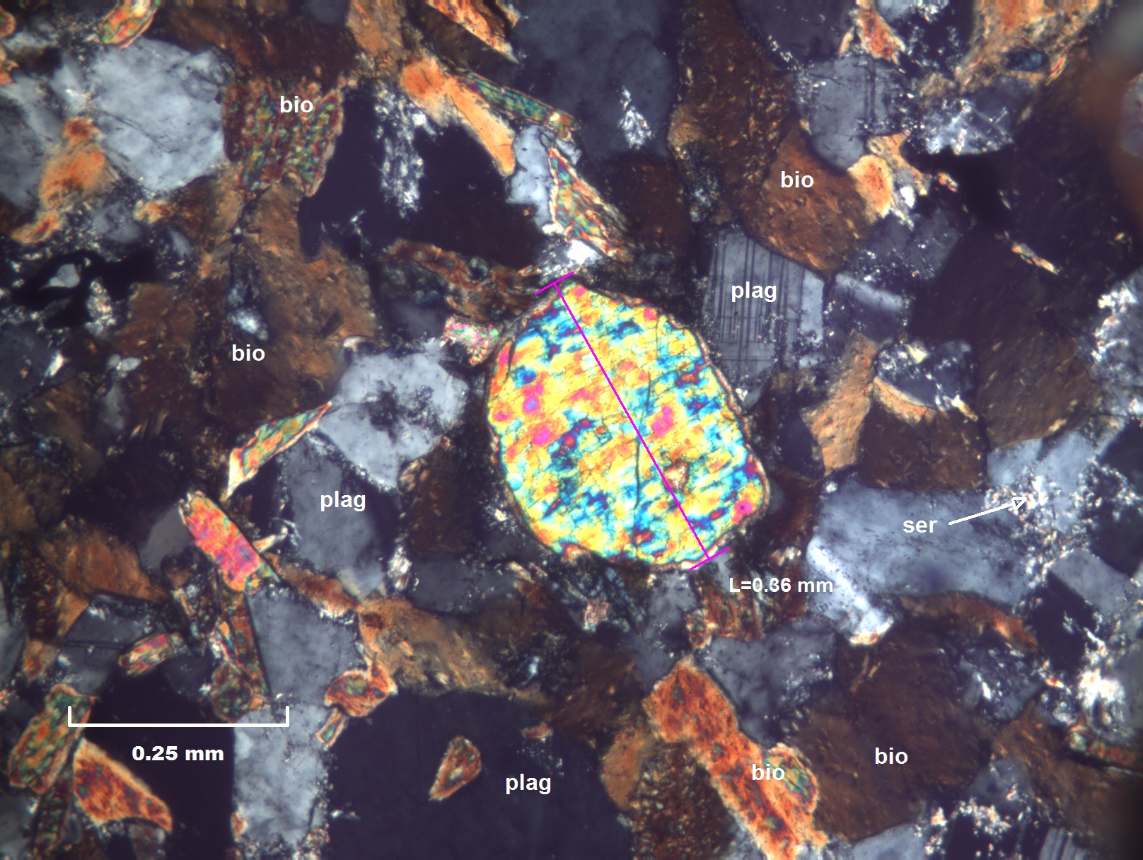
Zircon in Biotite Plagioclase Gneiss
10x Magnification, Crossed polarizers, 8ms Exposure, 2x Gain, 1.5 Gamma (Photo: Terry Crump)
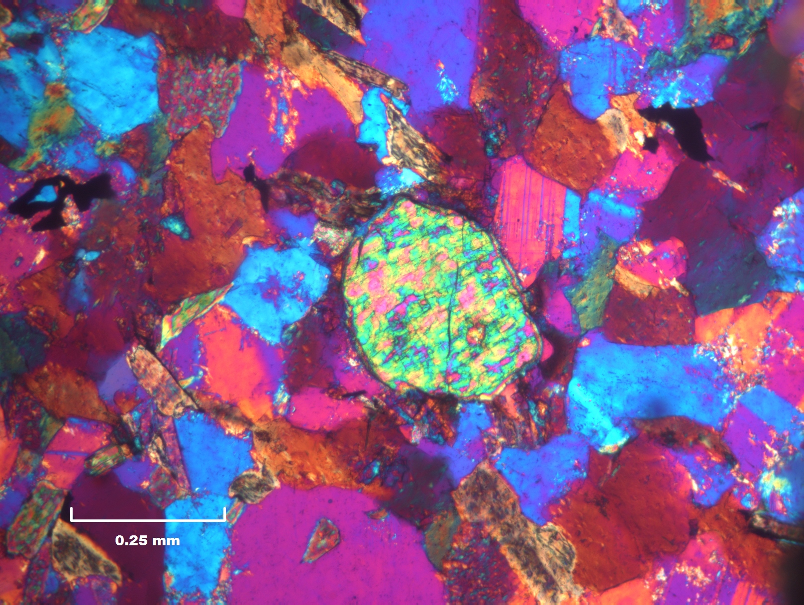
Zircon in Biotite Plagioclase Gneiss
10x Magnification, Crossed polarizers, 8ms Exposure, 2x Gain, 1.5 Gamma (Photo: Terry Crump)
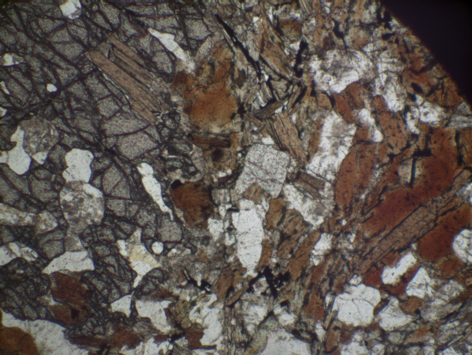
Biotite-Plagioclase Gneiss
4x Magnification, Plain polarized light, 1.3ms Exposure, 1.5x Gain, 1.8 Gamma (Photo: Terry Crump)
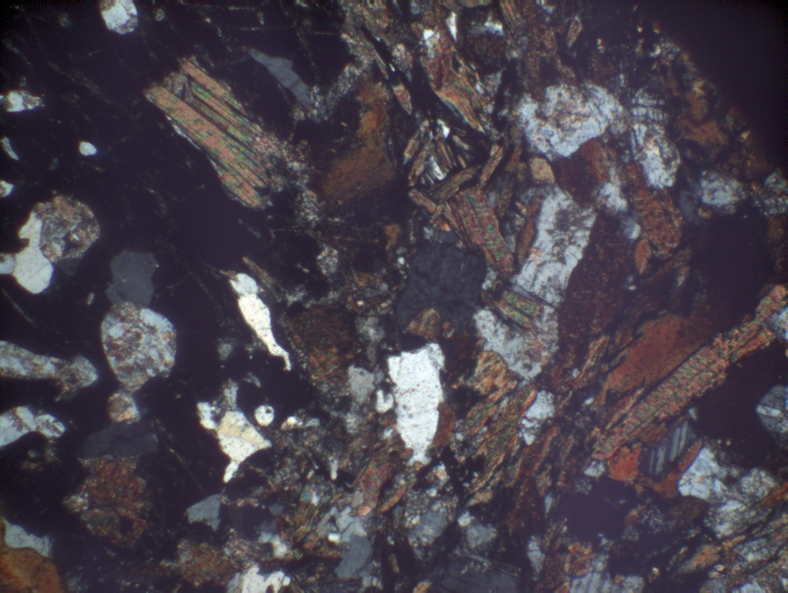
Biotite-Plagioclase Gneiss
4x Magnification, Crossed polarizers, 3ms Exposure, 2x Gain, 2.25 Gamma (Photo: Terry Crump)
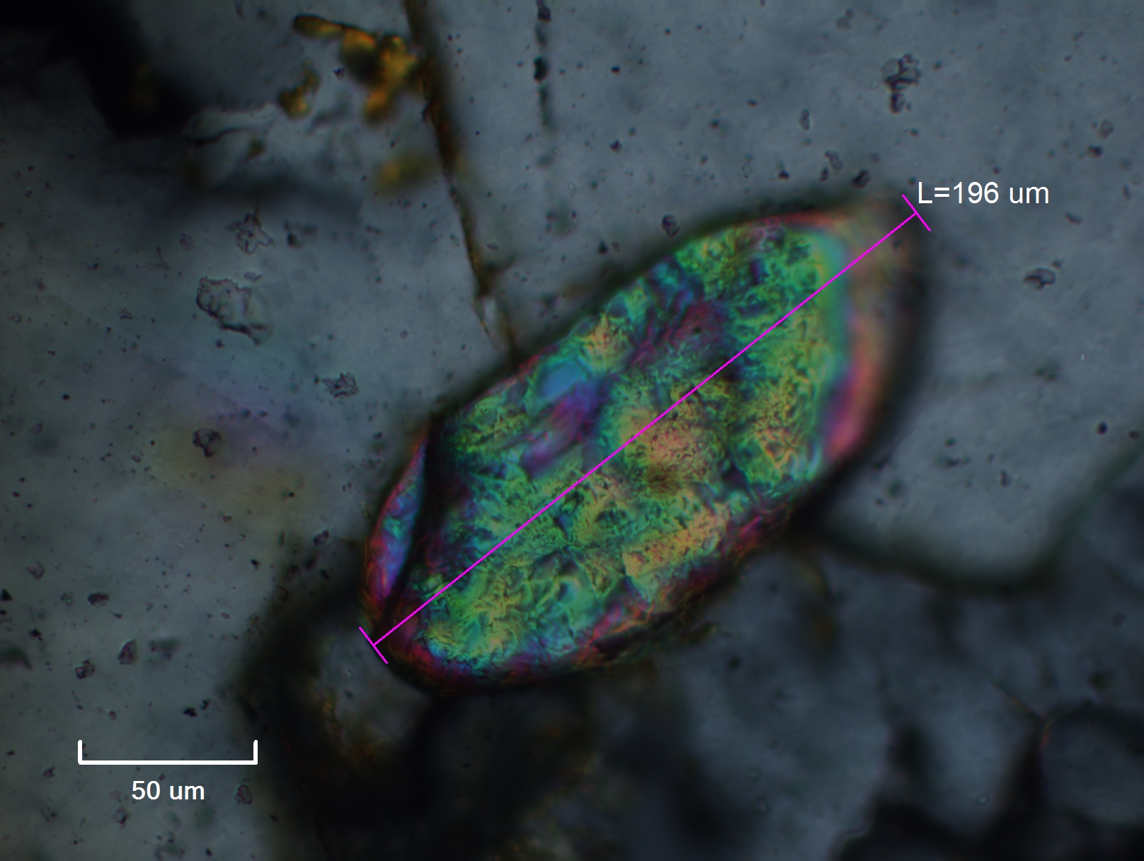
Zircon Crystal
40x Objective, Crossed polarizers, fluorescent source, 1 Gamma (Photo: Terry Crump)
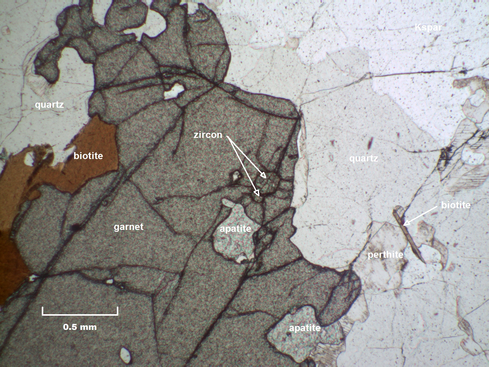
Garnet with Biotite, Apatite, and Zircon
4x Objective, Polarized light, 1.3.ms Exposure, 1.7x Gain, 1.7 Gamma (Photo: Terry Crump)
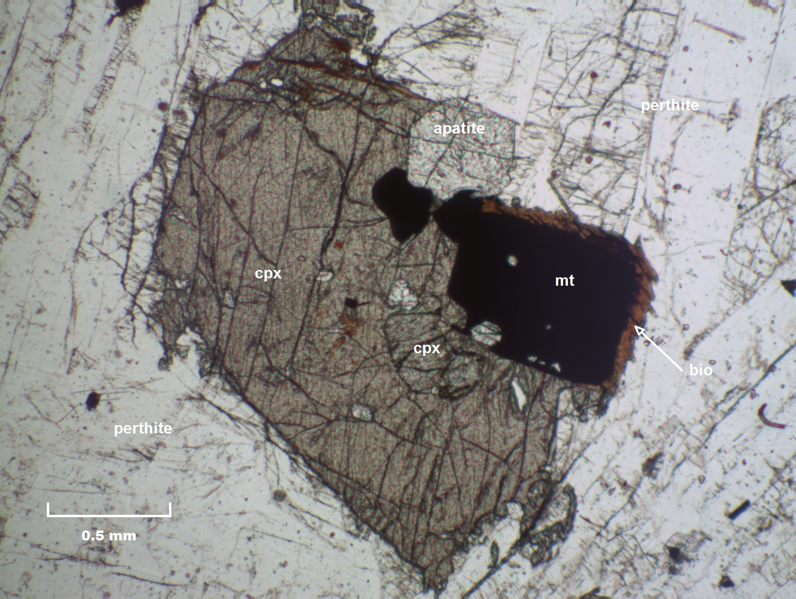
CPX Grain with Apatite, Magnetite, and Biotite
4x Objective, Polarized Light, 1.5ms Exposure, 1.5x Gain, 1.7 Gamma (Photo: Terry Crump)
Twinned CPX with Apatite, Magnetite, and Biotite
4x Objective, Crossed Polarizers, 10ms Exposure, 1.7x Gain, 1.8 Gamma (Photo: Terry Crump)
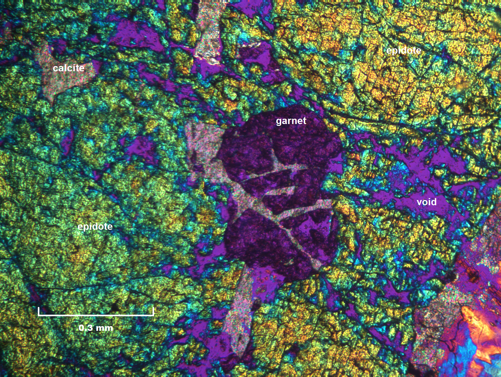
False color image of fractured garnet inclusion with calcite fracture filling in epidote
10x Objective, 20.ms Exposure, 2x Gain, 1.5 Gamma (Photo: Terry Crump)
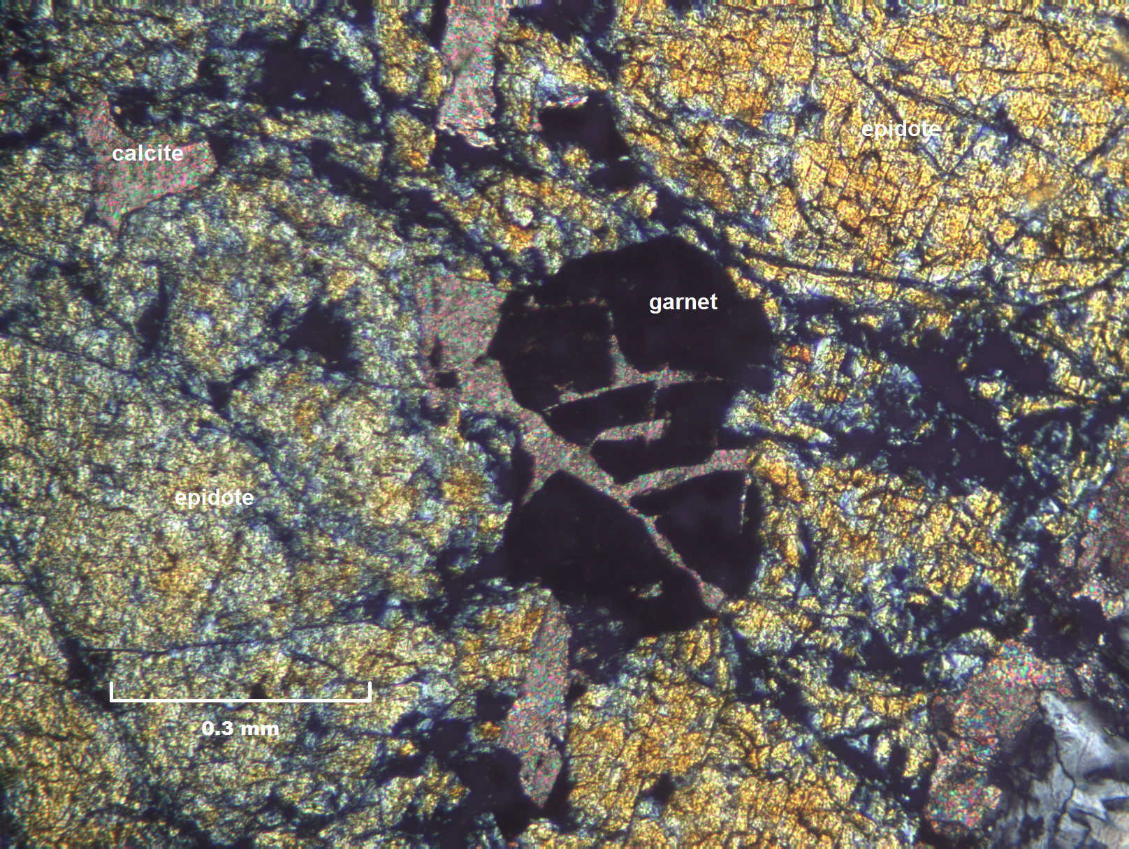
Fractured garnet inclusion with calcite fracture filling in epidote
10x Objective, 16ms Exposure, 2x Gain, 2 Gamma (Photo: Terry Crump)
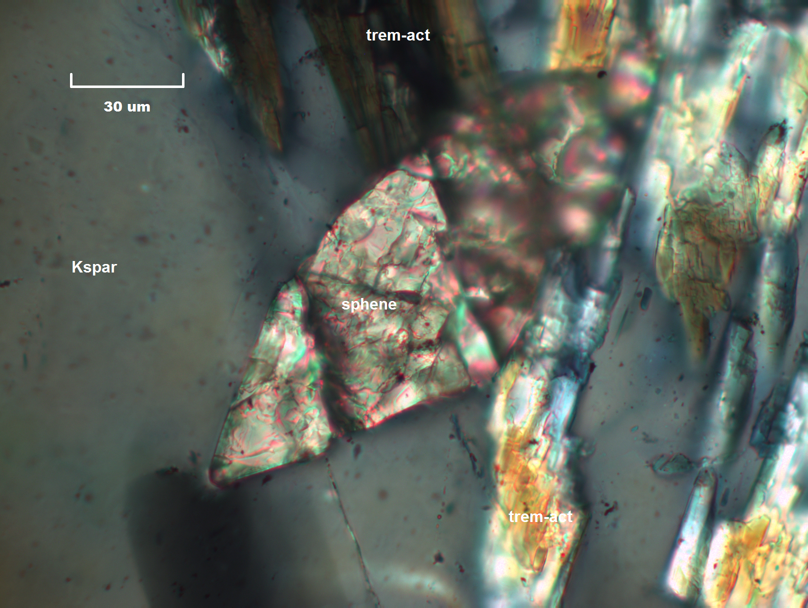
Sphene crystal with characteristic axehead shape in potassium feldspar and tremolite-actinolite
60x Objective, 50ms Exposure, 2x Gain, 1.2 Gamma (Photo: Terry Crump)
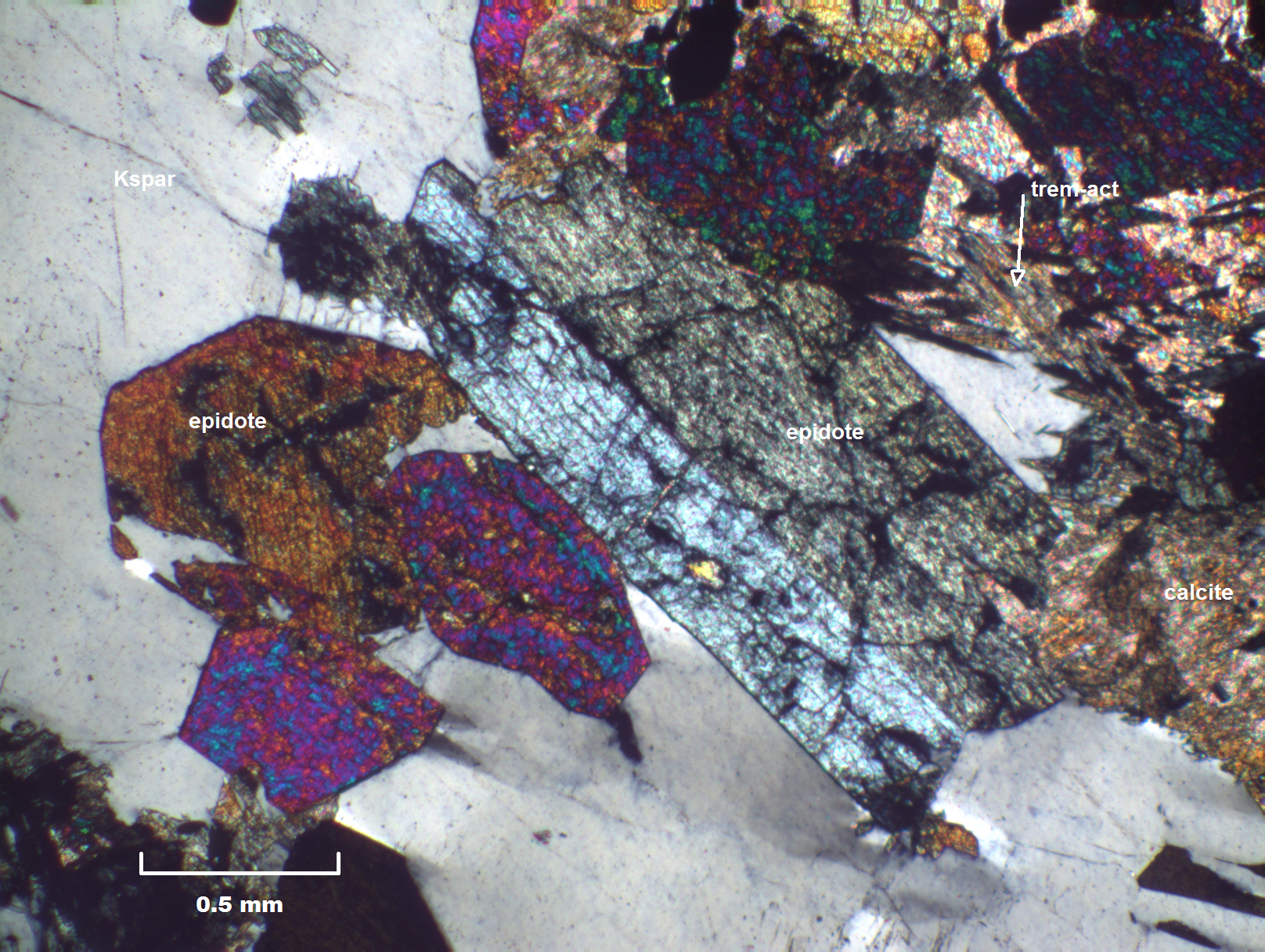
Twinned epidote accompanied by other epidote grains, calcite, tremolite-actinolite in potassium feldspar
4x Objective, 6ms Exposure, 2x Gain, 1.5 Gamma (Photo: Terry Crump)
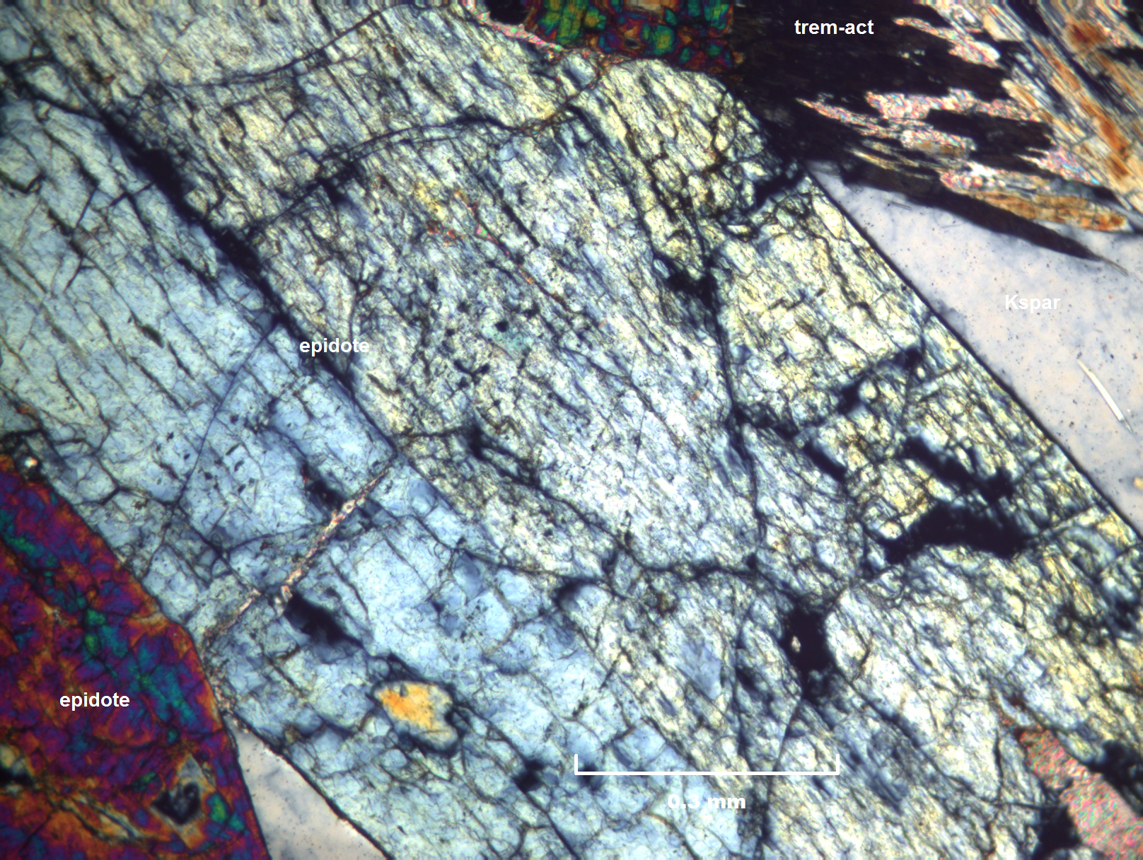
Twinned epidote showing single cleavage trending NW parallel to the long axis of the grain
10x Objective, 5ms Exposure, 2x Gain, 1.3 Gamma (Photo: Terry Crump)
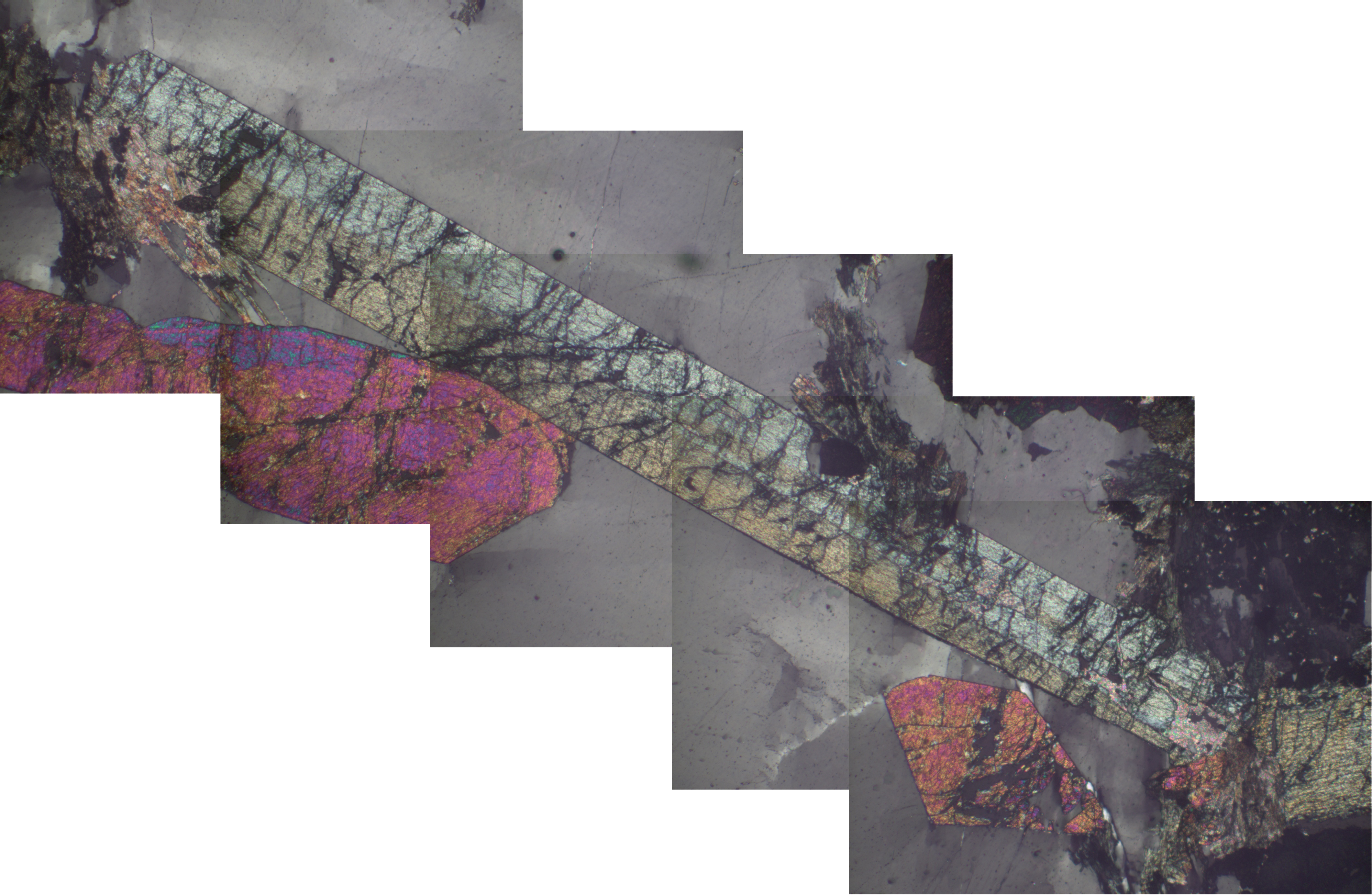
Panorama of epidote crystal collected at the Calvert mine in Beaverhead County, Montana
(Photo: Terry Crump)
