|
| |
|

PRODUCT SPOTLIGHTNEW RELEASE: INFINITY-EPHigh-Speed, Digital Microscopy Camera Designed for Electrophysiology
To learn more about the product click here. To find out how to order your camera: [email protected]
NEW SCIENTIFIC VIDEO"NEED A CAMERA FOR YOUR MICROSCOPE? CHOOSE LUMENERA'S INFINITY MICROSCOPY CAMERAS"
Lumenera's INFINITY cameras are compatible with any microscope, come with image analysis software, 4-year warranty, and exceptional customer support. Need help selecting a microscope camera? Learn more by watching our newest video.
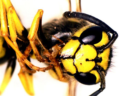
STUDENT PHOTO CONTESTANNOUNCING THE LUMENERA-BIOBUS STUDENT "CELL-FIE" PHOTO CONTEST WINNERAt the start of the September 2015 school year, Lumenera donated seven of their high-performance, research grade INFINITY microscopy cameras to the BioBus, a mobile microscope laboratory based in New York City that gives students the opportunity for hands-on scientific exploration and aims to provide children with access to modern science curricula and inspire disadvantaged youth to pursue careers in science. As part of this initiative, Lumenera and BioBus held the "Student Cell-fie Image Contest," where the best image taken all school year wins its class the Grand Prize of a $500 back-to-school supply shopping spree and a microscope to benefit the entire class. After choosing the top five best images submitted and opening up the final decision to the public in the form of an online poll, Lumenera and BioBus awarded the Grand Prize to the students at P.S. 196 in Bronx, New York for their close-up photo of a wasp.
FEATURED WHITE PAPERDIGITAL PATHOLOGY: A PRIMER FOR MICROSCOPY CAMERA SELECTION
Selecting the proper imaging equipment is one of the most important elements a pathologist needs to take into consideration when using a digital system. To produce a high quality pathology image, one must have a digital camera that can dependably and accurately reproduce what a pathologist sees in the eye piece of their microscope. Excellent color, high dynamic range and high resolution are the key elements that comprise a high quality pathology image. It is also important that pathologists can detect features in dark areas (which means the camera must have high sensitivity), that the camera doesn't add unwanted artifacts to the image (has low noise), and can be easily connected to a computer (via a USB interface, for example). This paper reviews the fundamentals of imaging within the field of pathology and what should be taken into consideration when selecting a digital microscopy camera.
FEATURED WEBINARCOLOR IMAGE QUALITY CONTROL IN MICROSCOPYLumenera recently sponsored the Bitesize Bio Webinar: "Color Image Quality Control in Microscopy"
Color imaging is everywhere today and is often taken for granted. Images are instantly captured on cell phones, tablets, web cams, and microscopes. Pictures are shared with friends and colleagues even faster. We thrive on instant gratification. For some of us, our images are simply a snapshot in time- a selfie in front of a landmark, an amazing meal presentation, a pic showing viable cells in culture. But for others, images need to be masterpieces, expressing not just artistry but scientific discovery. Regardless of your philosophy, we are at the mercy of the technology: cameras AND software. So as scientists, how do we ensure that we capture and communicate images that are worthy of our research? In this webinar, you'll learn about how color in images goes awry, and what you can do about it. And there's a twist at the end about an often overlooked villain in this whole scheme. Click here to watch a recorded copy of the webinar.
IN THE NEWSLUMENERA FEATURED IN THE OTTAWA BUSINESS JOURNAL"Lumenera hopes camera donation sparks 'passion for science' among students"
featured Blogs"SELECTING THE RIGHT COUPLER FOR YOUR MICROSCOPE CAMERA"
Read more here. "BACK TO SCHOOL WITH LUMENERA"
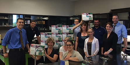
This school year, Lumenera donated 48 INFINITY microscope cameras to high schools across the Ottawa area- the community in which we operate and proudly support. By giving students access to the type of research-grade equipment that you would find in post-secondary institutions and professional research labs, Lumenera aims to foster a passion for science and inspire them at an early age to consider a career in the field. Read more here. Read more blog posts from Lumenera here. EVENTSWHERE WE'VE BEENLumenera at Neuroscience 2016
At Neuroscience 2016, Lumenera held two live demonstrations at their booth. In the first demo, we showcased our new, ultra-sensitive INFINITY3S-1UR in order to demonstrate its incredible fluorescence capabilities. The second demo featured our high speed 6 megapixel INFINITY3-6UR camera, exhibiting its amazing color reproduction and high resolution.
Lumenera at the Scottish Microscopy Meeting 2016
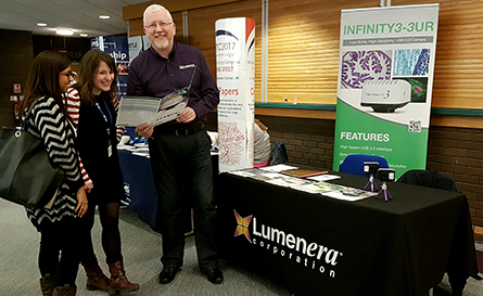
At the end of November, our European Director of Channel Sales, Patrick Cullen, attended the Scottish Microscopy Group's Annual Symposium to promote our high performance INFINITY microscopy cameras
UPCOMING EVENTSLight Microscopy Facility Managers Meeting 2017 Lumenera will be speaking SPIE Photonics West 2017 Lumenera will be exhibiting USCAP 2017 Annual Meeting Lumenera will be exhibiting Check out our Event page to see where we will be in 2017! CORPORATE UPDATECAREERS AT LUMENERALumenera was recognized two years in a row as one of the National Capital Region's Top 25 Employers, and is proud to offer a positive and rewarding workplace for our valued employees. We are currently looking to fill the following positions: Director of Product Management
LUMENERA'S HOLIDAY HOURSDecember 26th: Closed
|
In this issueConnect with us
supportcollateralContact Us
| |||||
|
Lumenera also distributes an Industrial e-newsletter. Send us an e-mail if you would like to automatically receive information on our other divisions. Lumenera Corporation, 7 Capella Crt., Ottawa, Ontario, Canada, K2E 8A7 Phone: 1-613-736-4077 Fax: 1-613-736-4071 If you prefer not to receive emails from Lumenera, or if you've changed your email address, please contact us at [email protected]. |
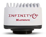 Lumenera's INFINITY-EP digital camera is a cost-effective solution with excellent near IR sensitivity and responsivity. This camera produces crisp, incredibly low noise images and videos are delivered at high frame rates with zero lag. Lumenera's Advanced Thermal Management Technology (ATMT) eliminates dark current noise, providing high-contrast imaging that facilitates the accurate placement of electrodes in tissue samples for electrophysiology applications.
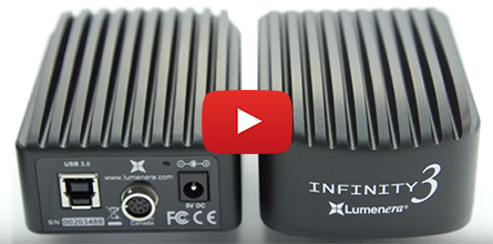
 Most pathologists today use digital systems within their practices and research projects to communicate, store data, and analyze images. Whether it's within clinical, forensic, surgical, or other branches of pathology, communication and consultation among specialists is greatly improved by the use of digital systems and enables faster diagnosis for patients.
  Overview: Mark Clymer, an expert in microscopy imaging, will guide you through the complicated area of color reproduction in microscopy. He will offer advice and tips on how to get the most from your microscopy camera.
 "Ottawa-based Lumenera Corporation has donated 48 research-grade microscope cameras to local high schools, the company announced on Tuesday. The maker of high-performance and custom digital cameras said it hopes the donation will help cultivate an interest in science among high school students."
 Selecting a suitable coupler to connect a camera to a microscope is an important step in the instrument's configuration. Another term for a coupler is a C-mount adapter, and the terms are often used interchangeably. When selecting a coupler, several factors must be considered in order to maximize the camera's field of view, without compromising image quality.